Magnetism in two-dimensional materials is difficult to characterize because the materials’ extreme thinness renders conventional techniques ineffective. Researchers in Australia, Russia and China have now used a new method called wide-field nitrogen-vacancy (NV) microscopy to measure the magnetic strength of vanadium triiodide (VI3), a 2D material that is known to be strongly ferromagnetic in its bulk, 3D form. The technique could also be used to study other 2D magnetic materials – including some possible building blocks for future energy-efficient electronic devices.
An NV microscope is a recently-developed instrument that uses defects in diamond as sensitive probes of weak magnetic fields. It is particularly well-suited for analysing samples of van der Waals materials (that is, materials composed of atomically thin layers that interact with each other via weak van der Waals forces) because it enables researchers to image magnetic domains (microscopic regions in which all magnetic moments point in the same direction) in individual flakes of the material with sub-micron resolution.
NV centres as detectors of weak magnetic fields
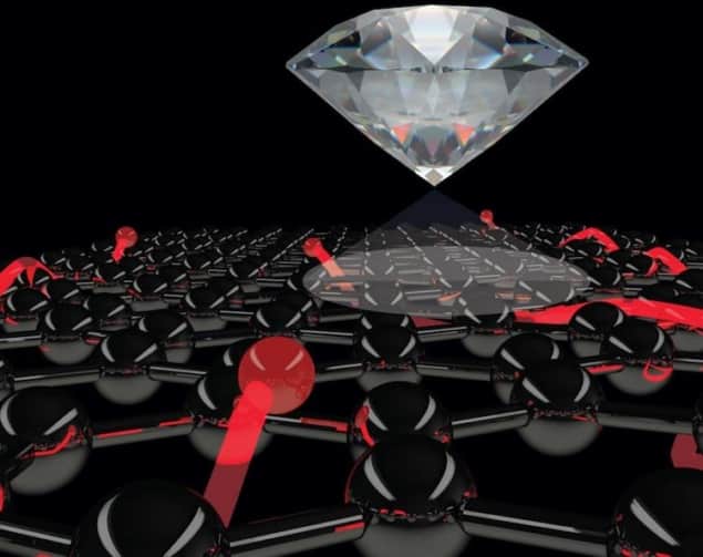
In the present study, researchers led by Lloyd Hollenberg of the University of Melbourne used an NV microscope made from a diamond substrate with a surface layer of defects. These defects are known as NV centres, and they occur when adjacent carbon atoms in the diamond lattice are replaced with a nitrogen atom and an empty lattice site. The nitrogen has an extra electron that remains unpaired, and therefore behaves as an isolated spin that can be up, down or a superposition of the two. The state of this spin can be probed by illuminating the diamond with laser light and recording the intensity and “spin-active” frequencies of the fluorescence emitted.
Because NV centres are naturally isolated from their surroundings, the spin state of their electrons is not immediately affected by surrounding thermal fluctuations. They can therefore be used to detect the very weak magnetic fields stemming from nearby electronic or nuclear spins – making them highly sensitive probes of magnetic resonance, capable of monitoring local spin changes in a material over distances of a few tens of nanometres.
Measuring magnetic field behaviour
In their experiments, Hollenberg and colleagues placed their sample of VI3 on top of the NV microscope’s defect layer, excited the NV centres with a laser and used a camera to image the resulting fluorescence. By sweeping the frequency of an applied microwave field across the sample (which they placed in a cryostat so that they could repeat the measurements at temperatures ranging from 4 to 300 K), they obtained what is known as an optically-detected magnetic resonance spectrum for their sample.
When the researchers applied a magnetic field of 0.5 to 1 Tesla perpendicular to their samples (the +z direction) at a temperature of 5 K, they observed complete and abrupt switching of the magnetic field direction in most flakes, with no intermediate partially-reversed state. This ferromagnetism persists down to two atomic layers, while the switching remains abrupt up to 50 K, which is VI3’s Curie temperature (that is, the temperature at which the bulk material loses its permanent magnetism).
VI3 is a nucleation-type hard ferromagnet
In hard magnetic materials like VI3, the direction-switching process depends on one of two mechanisms: nucleation or pinning of domain walls. In bulk materials, these mechanisms can normally be distinguished by their initial magnetization curves. These curves are produced by placing a sample of a magnetic material in a magnetic field and measuring how its magnetization increases.
In a nucleation-type magnet, the domain walls move freely. In a pinning-type magnet, as the name suggests, they are constantly being trapped. To determine which type of mechanism was at play in ultrathin VI3, Hollenberg and colleagues applied short magnetic pulses (around 10 nanoseconds long) in the -z direction to samples that had initially been magnetized in the +z direction and imaged the magnetization in a low magnet field after each pulse.
The results obtained suggest that ultrathin VI3 is a nucleation-type hard ferromagnet. However, the researchers also found that the magnetic strength of 2D VI3 is roughly half that of its 3D counterpart. “This was a bit of a surprise, and we are currently trying to understand why the magnetisation is weaker in 2D, which will be important for applications,” says team member Jean-Philippe Tetienne.
Diamond sensors probe matter at high pressures
According to Artem Oganov of the Skolkovo Institute of Science and Technology in Moscow, the group’s work could lead to new technologies. “Just a few years ago, scientists doubted that two-dimensional-magnets are possible at all,” says Oganov, who was also part of the research team. “With the discovery of two-dimensional ferromagnetic VI3, a new exciting class of materials emerged. New classes of material always mean that new technologies will appear, both for studying such materials and harnessing their properties.”
Members of the team, who include researchers from the University of Basel, RMIT University, Nanjing University of Posts and Telecommunications, Moscow Institute of Physics and Technology, Northwestern Polytechnical University, and Renmin University of China, say they now plan to use their NV microscope to study other 2D magnetic materials. They report their work in Advanced Materials.


