Proton minibeam radiotherapy (pMBRT) is an innovative new cancer treatment that uses an array of narrow proton beams to create alternating regions of high and low dose, before converging to a homogeneous dose distribution within the target volume. Numerous studies in small animals have shown that this spatially modulated dose distribution can destroy tumours while sparing healthy tissue and reducing side effects.
To implement pMBRT in the clinic, it’s important to first assess the impact of organ motion during treatment, which could lead to overlapping of the dose peaks and valleys, and potentially diminish the sparing effect. With this aim, a team at Institut Curie in France has developed a Monte Carlo (MC)-based framework to quantify the dose distribution for realistic clinical treatments, reporting their findings in Physics in Medicine & Biology.
To deliver pMBRT, a multi-slit collimator is used to create narrow beams, typically 0.3 to 1 mm wide, spaced 2 to 6 mm apart. As this collimator blocks much of the initial proton beam, longer treatments are needed to deliver the prescribed dose, increasing the likelihood of organ motion.
“Given the longer irradiation times, possible patient movement could deteriorate the heterogeneous spatial dose distribution of pMBRT,” explains senior author Ludovic De Marzi. “This is all the more problematic because it is this heterogeneous distribution that is believed to be responsible for the technique’s biological sparing effect.”
The dose reconstruction workflow
De Marzi and colleagues developed a 4D dose calculation workflow based on time-resolved MC simulations to determine how motion affects pMBRT dose distribution. The tool requires two main inputs: a 4D CT dataset capturing the patient’s anatomy and breathing motion over one respiratory cycle; and a proton pencil-beam scanning (PBS) treatment plan created on a reference CT.
To validate their workflow, the researchers irradiated a thoracic motion phantom with a conventional PBS plan and compared the measured dose with the simulations. For a 3%/3 mm gamma index (a standard quality check tool), the mean pass rate for nine phantom irradiations was 97.3%, confirming that the calculation performs reasonably well for 4D dose modelling in conventional PBS.
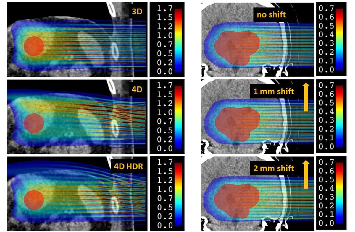
They then used the tool to assess the impact of motion in representative clinical cases, first examining a thoracic treatment with large breathing movements. They calculated 4D dose distributions for three scenarios: 3D pMBRT without motion; 4D pMBRT with 20 mm breathing motion; and 4D high-dose rate pMBRT, with the instantaneous dose rate increased tenfold to reduce beam-on time by a factor of ten.
The calculated dose distributions showed that breathing motion introduced significant variations in the patterns of peaks and valleys, impacting a crucial parameter: the peak-to-valley dose ratio (PVDR). At a depth of 5 mm, for example, the mean PVDR dropped from 11.9 for the 3D scenario to 6.7 in 4D. The high-dose rate scheme slightly mitigated this degradation, but not significantly.
“The PVDR is an index that takes into account both doses in the valleys (which are likely correlated with the biological sparing effect on healthy tissue) and the maximum delivered doses (which are likely responsible for the anti-tumour effect),” De Marzi explains. “It can also be used to help optimize the 3D dose distribution of the treatment plan.”
Breathing motion also degraded the dose delivered to the tumour, with the mean target dose and D95% (the minimum dose received by 95% of the target volume) reduced by 30% and 35%, respectively, for 4D pMBRT. Nearby organs-at-risk (OARs) were also impacted, with a decrease in dose to the stomach and a significant increase in mean dose to the spleen for the 4D cases.
The researchers next examined an intracranial treatment. While there is no intra-organ motion in this case, the patient’s head undergoes small rigid translations – generally limited to less than 1 mm using thermoplastic masks. As such, they modelled patient motion as a simple continuous 1 or 2 mm translation.
Motion did not significantly impact the mean dose or D95% for the target or OARs. A 1 mm shift was, however, enough to degrade the peak–valley patterns, reducing the mean PVDR by 10% from the static case (at 15 mm depth). For a 2 mm shift, this increased to a 24% reduction in mean PVDR.
A valuable tool
The researchers conclude that their 4D dose reconstruction workflow provides a useful tool for assessing interplay effects caused by organ motion during pMBRT. Without a specific motion management strategy, only intracranial treatment with movements below 1 mm was robust to interplay effects. A similar workflow could prove valuable for estimating the sparing effect of pMBRT while accounting for patient motion and adapting treatment plans accordingly.
Proton minibeams could improve treatment of cancer metastases

Looking forward, it may be possible to minimize delivery times by combining pMBRT with the ultrahigh-dose rate delivery used for FLASH treatments.
“This idea is appealing because it could both solve the problem of reduced dose rate in pMBRT and add an additional biological effect (FLASH) to further spare healthy tissue,” says De Marzi. “However, its feasibility remains to be demonstrated, as does the value of developing a technique that combines such considerable complexities. A simple increase in dose rate – without going as far as FLASH – would already be of interest.”
In their next step towards clinical application, De Marzi and colleagues are working to validate calculation tools and measurement procedures tailored to this pMBRT technique. “Ones that are fast, accurate and usable by clinicians,” he says.