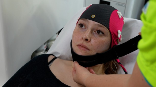
A system that recognizes the distinctive electrical signals in the brain that are associated with large ischaemic strokes has been developed by Jonathan Coutinho at the University of Amsterdam and colleagues in the Netherlands. The portable system has been used successfully in ambulances and with further improvements, the team says that the technique could find widespread use.
An ischaemic stroke is a serious condition that is triggered when a blood clot blocks the flow of blood to part of the brain. Ischaemic strokes account for about 85% of strokes in the UK and are treated using clot-busting medication, or in the case of large-vessel occlusions, (LVOs), the mechanical removal of the clot. But time is of the essence and treatment must start as soon as possible to minimize damage to the brain.
Today, medical imaging techniques like X-ray computed tomography (CT) and magnetic resonance imaging (MRI) are used to determine the type and severity of stroke a patient is experiencing.
The right hospital
“When it comes to stroke, time is literally brain,” Coutinho explains. “The sooner we start the right treatment, the better the outcome. If the diagnosis is already clear in the ambulance, the patient can be routed directly to the right hospital, which saves valuable time.”
Unfortunately, however, mobile facilities available today are not good enough to reliably diagnose strokes before patients reach the hospital. Now, Coutinho’s team have developed a new approach that involves monitoring patients’ brainwaves.
Brainwaves are rhythmic patterns in electrical activity caused by the synchronized oscillations of neurons in the brain. Their frequencies are closely tied with different states of consciousness and mental activity. In patients suffering from brain disorders, the synchronization between neurons can be thrown out of balance, altering the amplitudes and frequencies of the brainwaves produced. This is true for stokes because cutting off blood flow to a part of the brain affects the production of brainwaves there.
In some cases, these changes can be monitored using electroencephalography (EEG). This is a well-established technique whereby the subject wears a cap with holes for electrodes that contact the scalp and monitor the brain’s electrical signals in real time.
Today, EEG is mostly used to monitor the brainwaves of patients with epilepsy. Yet in their study, Coutinho and colleagues altered the cap’s design to detected signals unique to patients experiencing stroke symptoms. Their approach allowed them to determine whether or not a stroke is ischemic, while also indicating the size of the blocked blood vessel.
Very good news
“Our research shows that the brainwave cap can recognize patients with large ischemic strokes with great accuracy,” Coutinho explains. “This is very good news, because the cap can ultimately save lives by routing these patients directly to the right hospital.”
Portable MRI diagnoses stroke at the patient bedside
The team tested their brainwave cap in 12 ambulances in the Netherlands. Over four years, they collected data from over 400 patients – identifying LVO ischemic strokes with an impressive accuracy.
“This study shows that the brainwave cap performs well in an ambulance setting,” says Coutinho. “For example, with the measurements of the cap, we can distinguish between a large or small ischemic stroke.”
For now, further improvements will be needed before the team’s brainwave cap is approved for use in ambulances. Yet based on their early results, the researchers are confident in its potential to save the lives of many stroke patients, and to help minimize the risk of permanent brain damage.
The research is described in Neurology.


