Scientists have known for almost half a century that biological materials generate a voltage when they are exposed to a mechanical force. Now a team at the Oak Ridge National Laboratory and North Carolina State University has exploited this piezoelectric effect to produce the most detailed images of the internal structure of human teeth (S V Kalinin et al. 2005 arXiv.org/abs/cond-mat/0504232). The technique, which is known as piezoresponse force microscopy, could be used to image a wide range of biomaterials on scales of less than 10 nanometres.

Piezoelectricity is an intrinsic property of biological systems and is most pronounced in biomaterials that contain arrays of proteins or polysaccharides. The surface layer of a tooth is made of enamel, which consists mainly of crystals of a molecule called hydroxyapatite and a small amount of organic material — mostly protein — that is concentrated in the lower sub-layers of enamel. The inner layer of tooth, called dentin, contains a significantly higher fraction of collagen, which is the most abundant protein in nature. A team led by Sergei Kalinin of Oak Ridge and Alexei Gruverman at North Carolina State built up a detailed picture of a tooth that had been cross-sectioned along its length by measuring the intensity of the piezoelectric signal at different positions.
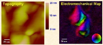
The team found that the enamel layer contains several isolated piezoelectric regions that are as small as 50 nanometres across, and that the dentin layer contains larger piezoelectric domains about 200 nanometres in size. This result is consistent with the fact that dentin contains a high density of collagen and enamel does not. In comparison, an optical image of the same tooth cannot provide this level of resolution. Moreover, the measurements revealed other details, such as the spiral shape of protein fibrils in dentin and how these fibrils are arranged antiparallel to each other (see figure).
“Our approach provides a new way to study structure and molecular organization in connective and calcified tissue such as teeth, bone and cartilage by monitoring their electromechanical properties,” says Kalinin. “Moreover, since piezoelectricity is ubiquitous in biological systems, the technique could be used to image almost any biomaterial.”


