A new twist on transmission electron microscopy (TEM) could enable the technique to unlock even more secrets on the nanoscale. Researchers in the US have produced a helical-shaped beam of electrons that could produce significantly higher-resolution images than is possible with conventional TEM, and it could be used to capture images of hard-to-spot bacteria and proteins.
TEMs work by firing a beam of electrons through a material and measuring how it absorbs and deflects the particles to build up an image of the sample. A microscope equipped with twisted electron beams should be able to produce images with even greater resolution thanks to the fact that the beams exchange large amounts of orbital angular momentum with the materials they interact with.
Twisted beams are already in used in optical microscopy, but it is much more difficult to twist beams of electrons. That is because electrons, like all other particles, have an associated wave whose wavelength is much shorter than that of light, so electron waves need to pass through much tinier structures to become twisted.
A special hologram
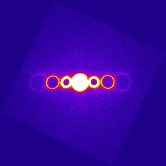
This has now been achieved by a group of researchers, including Ben McMorran of the National Institute of Standards and Technology (NIST), who fire electron beams through a specially designed hologram, which causes the beams to diffract. The diffraction created an ordinary plane wave beam, along with several helical-shaped beams, and the researchers were able to confirm the shape of beams and analyse how they evolve in time.
Although there are other ways to produce helical electron beams, the researchers say they used diffraction holograms because they more easily generate controllable beams with precise quantized large orbital momentum. The holograms were fabricated using a very finely focused ion beam to cut a pattern of extremely small slits just 20 nm across though a thin silicon membrane 30 nm thick. The free-standing silicon nitride structures are also quite mechanically robust and can withstand irradiation by the 300 keV electron beam in a TEM. And, they are small enough to be placed in the microscope without having to modify the instrument.
In addition to biological applications, the twisted electron beams could also be ideal for imaging magnetic materials because they can induce torques on charges in a sample by transferring angular momentum to them. “At its most fundamental, magnetism in a material is entirely due to the angular momentum of constituent charges, so being able to probe that using these beams will provide a new way to look at magnetic samples with unprecedented resolution,” said McMorran. “Quite recently another group confirmed this effect, which is very encouraging to us.”
Building on recent work
Indeed, a separate team based in Japan recently described an electron vortex beam produced by a different method and provided data on a single set of fringes showing that, while the electrons had spiral wavefronts, they were not single quantized orbital states. And a third group, based in Europe, described a similar technique to NIST’s but the holograms made were on the micro-scale as opposed to the nano.
“We made more complex, tinier holograms that enable us to achieve 10 times the separation angle between beams – important for applications – and 100 times the orbital angular momentum on electrons,” explains McMorran. “This is possible because each grating in our hologram produces multiple beams with higher diffraction orders containing proportionally larger amounts of angular momentum.”
The team is now working on ways to make the holograms even smaller. “We are taking a more detailed look at the fundamental properties of these helically shaped electron beams too, which is interesting stuff in itself. And to top it all, we are developing theory to understand all of this,” says McMorran.


