X-ray imaging is a valuable tool for studying biological systems, providing extremely high resolution as well as the ability to see within thick samples. The recent development of coherent X-ray sources has enabled the introduction of lensless imaging techniques, which eliminate the problem of creating suitable optics for X-ray microscopy.
Generation of the coherent X-ray radiation needed for lensless imaging generally requires large facilities such as synchrotrons or free-electron lasers. But in the extreme ultraviolet (EUV) spectral region (124–10 nm), coherent radiation can be created via high harmonic generation (HHG) using intense femtosecond lasers. Such HHG sources could, in principle, enable coherent EUV imaging to be performed in a small-scale laboratory.
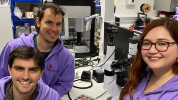
An international research team, led by Bill Brocklesby and Jeremy Frey at the University of Southampton, has now developed a laboratory-scale coherent EUV source from a femtosecond laser, and used it to create high-resolution images of lab-grown neurons. Such highly detailed images could have many potential applications in medicine and biology, including the study of neurodegenerative diseases.
Lose the lens
In lensless imaging, the object is illuminated with coherent radiation and the scattered radiation is collected onto a detector without requiring imaging optics. The image is created by algorithmic reconstruction of the phase of each pixel in the detected scatter pattern. In this study, the team employed ptychography – a type of lensless imaging in which the illumination is moved relative to the sample and multiple scatter patterns are recorded.
To test their approach, the researchers grew mouse hippocampal neuron samples on silicon nitride membranes in vitro. They first imaged the samples using an optical microscope. They then performed ptychographic imaging of small areas of the samples, using coherent 29 nm (43 eV) illumination produced by generating high harmonics from a pulsed femtosecond laser.
Ptychographic imaging with the EUV source created images that were quantitative in both amplitude and phase, with a lateral resolution of 80 nm and an axial sensitivity of approximately 0.8 nm (equivalent to a layer of protein). The high lateral resolution is due to the short wavelength and the accuracy of the phase retrieval algorithm, while the high axial sensitivity arises from the strong interaction of EUV radiation with the biological materials being imaged.
Comparison of an image taken using a phase-contrast visible light microscope with the EUV ptychographic image showed that the EUV image had much higher lateral resolution than its optical counterpart. For example, the EUV image showed fine “flared” structures emanating from thicker dendrites that were not seen in the optical image.

Fluorescence comparison
The researchers – who performed these studies at Southampton and the Artemis facility in the Rutherford Appleton Laboratory – also used EUV ptychography and conventional fluorescence microscopy to image neuron samples that were stained for immunofluorescence imaging. They observed that the EUV resolution was significantly better. EUV imaging was also more sensitive to thin structures in the neurons, identifying features of below 100 nm in width and only 10 nm thick, which could not be seen using fluorescence imaging.
The authors note that even with super-resolution fluorescence techniques, which can have lateral resolution smaller than the diffraction limit, fluorescence imaging of very thin structures will remain difficult, due to the extremely small quantities of fluorescent material present. They add that correlative EUV and fluorescence imaging allows the structural elements seen using EUV ptychography to be directly correlated to biological function.
Finally, the team examined the impact of the EUV radiation on the samples. Many other high-resolution techniques used for imaging biological samples, such as electron cryo-microscopy or hard X-ray microscopy, are limited by the damaging effects of the radiation. The 29 nm EUV radiation, however, did not damage the delicate neuron structure on the exposure timescales used for imaging.
“The ability to take detailed images of delicate biological structures like neurons without causing damage is very exciting, and to do it in the lab without using synchrotrons or other national facilities is a real innovation,” says Brocklesby. “Our way of imaging fills an important niche between imaging with light, which doesn’t provide the fine details we see, and things like electron microscopy, which require cryogenic cooling and careful sample preparation.”
The research is described in Science Advances.