The ability to track lung tumours during radiotherapy, without the use of markers, could make therapeutic dose delivery more effective and reduce exposure to healthy surrounding tissue. One option, the use of X-ray imaging, has been limited by its inability to adequately visualize a tumour overlapping bone. Researchers from Loyola University Medical Center have now shown that fast-kilovoltage (kV) switching dual-energy fluoroscopy can improve the accuracy of markerless tumour tracking when used with software that removes bone and enhances soft tissue (Med. Phys. 10.1002/mp.13573).
The research team previously evaluated sequential dual-energy imaging using the on-board imager of a linear accelerator (Med. Phys. 10.1118/1.4903892). This research revealed the efficacy of sequential dual-energy fluoroscopy, as well as its limitations, as it produced respiratory and organ motion artefacts. The team hypothesized that by rapidly alternating the X-ray tube potential at a high frame rate, such motion artefacts would be reduced and dual-energy imaging could be performed in real time.
For their latest study, the researchers employed a fast-kV switching fluoroscopy prototype developed at Varian Medical Systems that produced alternating 60 and 120 kVp X-rays. To simulate chest anatomy and tumour respiratory motion in the lung, they used a dynamic thorax motion phantom containing a 3D anthropomorphic spine and ribs and a spherical “tumour” capable of producing complex motion. They imaged five tumour inserts of 5 to 25 mm in diameter. The inserts were programmed to move in the phantom’s inferior–superior direction, perpendicular to the X-ray beam, to mimic respiratory motion.
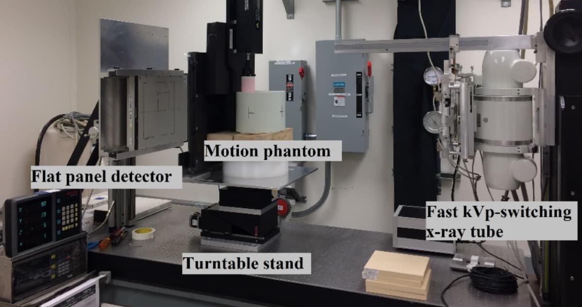
The researchers acquired images at 15 frames/s and created bone-subtracted images using internally developed software. They then calculated tracking success rate and accuracy in regions of the phantom where the target overlapped ribs and spine, to compare the performance of single-energy and dual-energy imaging methods.
The team also varied the CT slice thickness to evaluate and optimize template quality. They determined that a CT slice thickness of 0.75 mm resulted in the lowest tracking error for the smallest 5 mm target.
“The results of our study indicate that there may be a range of optimal slice thicknesses for each tumour size,” the researchers write. For single-energy imaging, the smallest CT slice thickness may be preferable for tumour diameters between 5 and 10 mm, while for larger tumours (15–25 mm) a CT slice thickness of less than 3 mm is desirable. For dual-energy imaging, a slice thickness of less than 3 mm would result in comparable tracking error for all targets below 25 mm in diameter.
“The most significant gains were observed for the smallest simulated tumours, the 5 and 10 mm diameter targets,” explains academic co-principal investigator John Roeske. “In particular, for the 5 mm target, there were cases where the motion was only tracked on a few frames using single-energy imaging. The addition of dual-energy imaging significantly improved the tracking success rate of this small target.”
The authors calculated the tracking accuracy for both single- and dual-energy imaging, using the mean absolute error between the predicted and expected location of the tracked tumour. With the exception of the 5 mm target, dual-energy imaging outperformed single-energy imaging in all simulated motion types. When the 5 mm tumour overlapped the simulated spine of the phantom, dual-energy tracking success rates for static, slow and fast motion were 24%, 38% and 54%, respectively, compared with 0%, 3% and 3% for single-energy tracking. In general, larger improvements were observed for tumours overlapping spine than those overlapping ribs.
The authors also note that higher peak-to-side lobe ratios and improved tracking accuracy of dual-energy over single-energy imaging may offer advantages in the clinical setting, where tumours may have varying densities and less than ideal shapes.
This research was a joint initiative between Loyola University Medical Center and a Varian research laboratory. Roeske tells Physics World that working with industry co-principal investigator Hassan Mostafavi, they are moving their studies from a benchtop system to a linac and have already implemented fast kV switching in Developer Mode of the TrueBeam️ radiotherapy system. They are also repeating a number of studies to validate their findings using the benchtop system. After they complete these studies, the goal will be to evaluate their approach, with IRB (institutional review board) approval, on a small cohort of patients.
When asked to speculate on when this markerless tumour tracking technology could be used in clinical settings, Roeske says that lead author Maksat Haytmyradov will present the initial characterization of fast-kV switching at this year’s ASTRO annual meeting.