“There are two fundamental issues with conventional MRI contrast agents,” explains Leif Schröder from the Leibniz-Forschungsinstitut für Molekulare Pharmakologie (FMP) in Berlin. “They are very insensitive and require high concentrations, and the gadolinium-containing ones impose a safety issue for certain patients.”
Along with collaborators from the California Institute of Technology (Caltech) led by Mikhail Shapiro, Schröder’s research team has developed a new type of contrast medium for MRI. This medium not only addresses the limitations of conventional contrast agents but also automatically adjusts to accommodate different amounts of hyperpolarized xenon gas (ACS Nano 10.1021/acsnano.8b04222).
The spins align
MRI is central to diagnosing and monitoring treatment of diseases. It creates images of the body by exposing water molecules in tissues to a strong magnetic field, avoiding potentially harmful radiation associated with other imaging techniques.
Contrast media, either injected or inhaled, are used to increase the sensitivity of MRI. Such media consist of contrast agents, and in some cases, targeting units that bind them to specific cellular disease sites. Agents can be detected indirectly through the water signal when they bind and exchange hydrogen atoms with those in water molecules.
This chemical exchange can be measured with greater sensitivity using hyperpolarized nuclei — an approach that has been applied to several noble gases, including xenon. Scientists can create hyperpolarized xenon gas using an optical pumping technique. This process uses laser light to pump electrons into higher energy levels and eventually align (or hyperpolarize) their spins. This polarization can be transferred onto the spin of nearby noble gas nuclei through spin exchange processes.
The most useful xenon isotope for MRI applications is 129Xe. Its prolonged hyperpolarized state can last up to several hours in gaseous form, and it can be used to image cavities in a porous sample, such as alveoli or gas flow within the lungs. Because xenon is soluble both in water and hydrophobic solvents, hyperpolarized 129Xe also can help doctors visualize various soft tissues.
However, as Schröder mentions, MRI requires a high concentration of molecules to generate a useable signal. Even using hyperpolarized xenon gas, the sensitivity of MRI remains low, meaning that many cellular biomarkers cannot be detected using current methods.
Swim bladders for xenon
The FMP–Caltech collaboration is working to improve MRI sensitivity by developing contrast media based on hyperpolarized xenon gas. Previously, they described a new class of contrast media that binds to xenon reversibly; however, how well these media could take up hyperpolarized xenon was unknown.
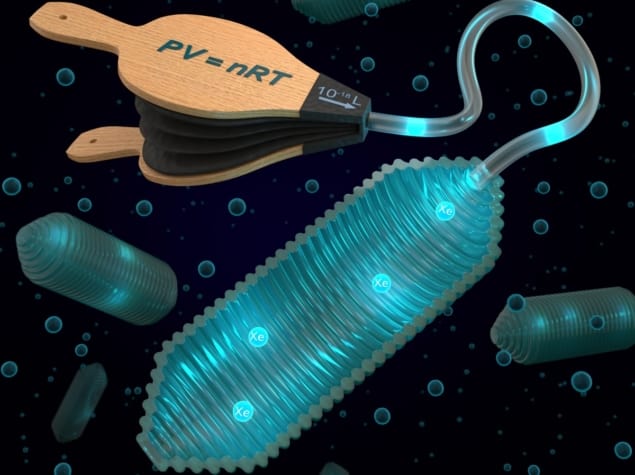
These new contrast media are hollow protein structures, or “gas vesicles”. Produced by certain bacteria, the gas vesicles function like the swim bladder of a fish, Schröder describes, allowing the bacteria to regulate their buoyancy in water.
New research by the FMP–Caltech team has demonstrated that the gas vesicles can adjust their influence on measured xenon signals according to the ideal gas law.
“The protein structures have a porous wall structure through which xenon can flow in and out. Unlike conventional contrast media, the gas vesicles always absorb a fixed portion of the xenon that is provided by the environment,” Schröder explains.
Therefore, the more xenon available, the more will be absorbed by the vesicles. MRI can take advantage of this accumulation and subsequent absorption.
The fraction of xenon gas that a patient inhales determines the fraction of xenon dissolved in their blood. Xenon encountering the gas vesicles in tissue will partition into the vesicles. Because much more xenon passes into the gas vesicles than with conventional contrast media, this improves both sensitivity and image contrast. This may also enable these new contrast media to identify disease markers occurring in low concentrations.
The future of gas vesicles for MRI
Schröder, Shapiro and their research teams have now produced the first MR images employing gas vesicles and hyperpolarized xenon gas. In the future, the researchers will employ gas vesicles to target different disease markers, such as binding to cancer cells or tracking immune cells. They also plan to quantify the improvements in sensitivity that can be achieved over conventional contrast agents.


