Gold nanoparticles (GNPs) show potential as CT contrast agents for imaging macrophages in areas of vascular inflammation, according to preclinical research from Japan. Such GNPs could also act as light absorbers in photothermal therapy, the research team report in Molecular Imaging and Biology. These findings could one day help in the diagnosis and treatment of vascular diseases such as atherosclerosis (plaque build-up in arteries) and abdominal aortic aneurysm (a swelling in the aorta).
Macrophages are specialized white blood cells involved in the detection and destruction of bacteria and other harmful organisms. They contribute to the progression of vascular inflammation, playing a key role in plaque formation, aneurysm progression and rupture. As such, macrophages could act as key imaging and therapeutic targets for vascular disease.
In an initial in vitro study, the researchers – from Tokyo Medical University, the University of Tsukuba and the AIST Nanomaterials Research Institute – incubated mouse macrophage cells with GNPs (50–200 µg/ml) for 24 hr and then scanned the cells with a micro-CT system. The CT attenuation values of macrophages incubated with GNPs were significantly higher than those without GNPs. The team note that GNP incubation did not decrease the viability of the macrophages.
Next, the researchers evaluated the use of intravenously injected GNPs for in vivo imaging of vascular inflammation in mice. They used ligation of the left common carotid artery to induce macrophage-rich atherosclerotic lesions in nine mice. The non-ligated right common carotid artery served as a control. Two weeks after the ligation, the team injected the mice with 10 or 20 mg of GNPs and scanned the animals with a micro-CT system, 24 and 48 hr after injection.
First author Hisanori Kosuge and colleagues report that the in vivo CT images exhibited contrast enhancement in both the diseased left and non-diseased right carotid arteries, at 24 and 48 h after GNP injection. Although the ratio of CT attenuation values in ligated versus non-ligated arteries was higher at 48 h than at 24 h, in both 10 and 20 mg groups, the difference was not statistically significant.
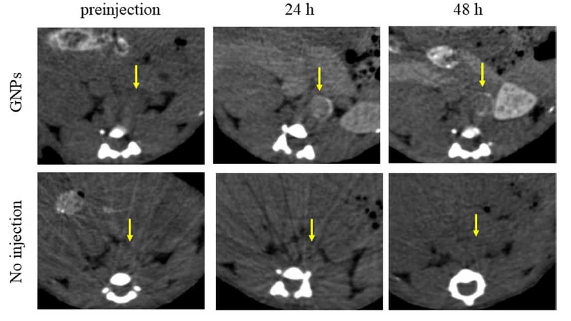
In a second in vivo study, the researchers induced abdominal aortic aneurysms in a group of male mice with deficient levels of apolipoprotein E, a protein involved in the metabolism of fats. They used ultrasound imaging to identify nine mice with aneurysms and injected five of them with 10 mg GNPs. After 24 and 48 hr, the researchers scanned all nine mice using micro-CT.
The CT images exhibited contrast enhancement in the perivascular area in mice injected with GNPs, while no enhancement was visible in non-injected mice. Ex vivo imaging of surgically exposed aortas confirmed that the CT attenuation values were significantly higher in mice injected with GNPs.
Therapeutic potential
The researchers also investigated the use of GNPs as light absorbers for photothermal therapy, by assessing the viability of GNP-incubated macrophages before and after exposure to pulsed near-infrared (NIR) laser light. They incubated macrophages with 0, 100 or 200 µg/ml of GNPs for 24 hr, and then exposed the cells to an 830 nm NIR laser. Low-intensity (178 or 196 mW) NIR laser light did not affect the viability of macrophages. However, high-intensity (400 or 437 mW) irradiation significantly reduced the viability of macrophages treated with GNPs (100 or 200 µg/ml) compared with those without GNPs.
Open-source algorithm predicts heart attack risk from chest CT scan
While the experiment showed potential for vascular inflammation therapy using a NIR laser, the mechanism of reduced viability was unclear. The researchers note that it may be difficult to reduce cell viability in vivo using the same laser conditions as in vitro, because blood flow through vessels leads to insufficient target heating. They suggest that further studies are required to examine the effects of laser irradiation.
The researchers are optimistic about their study findings. “In vivo CT imaging with GNPs successfully depicted experimental carotid atherosclerosis and abdominal aortic aneurysms, and NIR laser irradiation reduced the viability of macrophages incubated with GNPs,” they conclude. “Thus, GNPs have a strong potential for non-invasive CT imaging and therapy for vascular inflammation.”