
Computer-controlled 3D printing is now enabling the custom manufacture of many different products and structures, including tissue scaffolds that are designed to grow artificial tissues and organs in the lab. Researchers are busy exploring the performance of porous designs that contain different cells and growth factors, with the aim of repairing or replacing damaged parts of the body.
But the possibilities for this fast-evolving medical technique doesn’t stop there. Experts are also investigating whether the technology is compatible with plant cells, which could, for example, help to nurture active agents for pharmaceuticals, food and cosmetics.
Michael Gelinsky and his team at the Centre for Translational Bone, Joint and Soft Tissue Research at the University of Dresden in Germany have found that a bioink developed for printing human cells could be adapted to fabricate 3D plant cell cultures. “Having demonstrated bioprinting of human mesenchymal stroma cells with a novel and self-made alginate/methylcellulose bioink, we wanted to try other cell types to explore the applicability of this new blend,” explains Gelinsky.
Green bioprinting
Gelinsky and his team tested the concept by printing a cube-shaped mesh with a bioink loaded with basil cells. Reporting their results in the journal Biofabrication, the scientists observed that the majority of cells survived 3D plotting and cross-linking of the structure. What’s more, the embedded cells displayed high viability and metabolic activity during the investigated cultivation period of 20 days.
There are some fascinating applications to consider. The green bioprinted cubes could help developers to optimize the extraction of plant-based compounds, plus the work could come full circle and benefit the function of conventional scaffolds.
“As both plants and algae produce oxygen by photosynthesis, green bioprinting has the potential to keep mammalian cell cultures, or even tissues, alive,” says Gelinsky. “This feature could be of special interest for applications in space.”
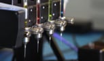
Additive manufacturing experience
Gelinsky’s group started using extrusion-based 3D printing for fabricating tissue scaffolds in 2010. Today, the group uses multiple print heads and concentric nozzles to combine a variety of biomaterials in a single structure.
This set-up allows the researchers to include stiffer biomaterials as support structures for the soft hydrogel elements of scaffold designs. In addition, concentric strands can be extruded with different drugs or growth factors loaded in the core and shell. The positioning of these active elements offers a dual-release profile, and varying the shell thickness and core composition provide ways of fine-tuning the release kinetics.
Considering tissue regeneration, this design feature paves the way for scaffolds that can release different groups of growth factors sequentially. In other words, it would enable a two-step treatment that could, for example, first reduce inflammation and then promote vascular ingrowth, as the group mentions in related work.
The precision and reproducibility of computer-controlled manufacturing methods could also benefit other aspects of scaffold development, including the translation of ideas from the lab to the clinic. “Bioprinting might help in standardizing and automating the fabrication of clinically applicable tissue engineering products,” Gelinsky points out.
The past, present and future of 3D bioprinting
Like other groups working in this field, Gelinsky’s team has strong links to the medical community, and its skill set extends throughout the university. “Biofabrication requires expertise in materials and knowledge of (stem) cell biology and cultivation, but it also demands engineering skills and an understanding of microfluidics, to list just some of the experience required,” he comments. “It’s a highly interdisciplinary field.”
The team posts regular updates on its biofabrication research on its Twitter page.
- Read our special collection “Frontiers in biofabrication” to learn more about the latest advances in tissue engineering. This article is one of a series of reports highlighting high-impact research published in the IOP Publishing journal Biofabrication.


