As the COVID-19 pandemic continues across the world, the wearing of face masks indoors has become a requirement to help reduce virus transmission. Facial coverings are also worn in MRI scanners during data acquisition to keep participants safe. However, it was unclear what impact this could have on measured brain signals. Now, researchers at Stanford University have investigated the effect of wearing a face mask on functional MRI (fMRI) signals during scanning. They describe their work in NeuroImage.
Functional MRI measures the blood-oxygen-level-dependent (BOLD) response of the brain due to changes in blood flow during activation. The BOLD response is sensitive to the concentrations of oxygen and carbon dioxide (CO2) in the blood. Wearing a facial covering mixes the expired and inspired air streams. As you breathe out CO2, this mixing increases the amount of inspired CO2, resulting in mild hypercapnia (elevated blood CO2 levels). This hypercapnia increases cerebral blood flow and therefore elevates the measured BOLD signal, resulting in greater contrast in the fMRI compared with that seen in the absence of hypercapnia.
The impact on BOLD signals
The Stanford team performed task-based neuroimaging studies using a “block design”, in which an activity is performed or a stimulation is given during an “on-window”. This is followed by an “off-window”, where the action or stimulation ceases.
In this study, the researchers ran two block designs simultaneously. The first involved a short 15-s sensory-motor task, which stimulated the brain’s auditory, visual and sensorimotor regions concurrently. Throughout the task, they delivered fresh air to the participant through a nasal cannula in on–off cycles of 90 s. They conducted the experiment twice with each participant, once with a surgical face mask and once without. The cannula manipulated the gas content of the inspired air during the mask-on state, preventing CO2 build-up; it had a minimal effect on CO2 levels when the mask was off.
The team also recorded end tidal CO2 (ETCO2) levels in a separate session outside of the MRI scanner, to measure the effect of the mask on hypercapnia for each participant.
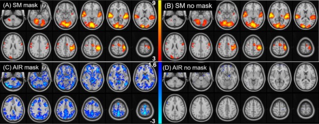
The researchers analysed data from eight healthy participants using a general linear model with two variables: one describing the sensory-motor task, and the other the nasal cannula air supply. The resulting group activation maps from the sensory-motor task, which indicate the areas of the brain that are active during the task on-window, showed no significant differences between the mask-on and mask-off states. These results demonstrate that task-activation can be reliably detected while the participant wears a mask.
The baseline BOLD signal, which the team analysed using the nasal cannula air cycles, showed a significant difference between the mask-on and mask-off states. The results demonstrated that the face mask induced an average baseline signal shift of 30.0%, with the grey matter across the brain showing an evident deactivation (observed via an increase in signal without the air supply) in the group activation maps. The measured ETCO2 showed an average increase of 7.4%, confirming the predicted rise in inspired CO2 concentration with mask use.
This topical study provides some clarity to the neuroimaging community regarding the impact of face masks on data collected throughout the pandemic. The insignificant difference measured between the task-activated signal with masks on or off supports the safe continuation of task-based fMRI studies in clinical and research settings while following mask regulations.


