Monitoring the healing of severe bone fractures is important because patients with such injuries are particularly susceptible to complications. Delayed bone healing that is not treated in a timely manner may cause severe pain and, ultimately, a loss in muscle strength and mobility.
The first few weeks following trauma influence whether the fracture will heal properly. The current solution is to wait until some evidence of successful healing is seen, such as the ability to bear weight. Despite over 40 years of scientific studies, monitoring non-union fractures is still a challenge. In fact, the remaining bottleneck in fracture care is the lack of clinical tools that enable direct, quantitative assessment of fracture healing and early diagnosis of non-union conditions.
Several approaches have been proposed to quantify fracture healing in long bones, ranging from mechanical vibrations to electrical impedance. Both in vitro and in vivo experiments have demonstrated the capabilities of different techniques to monitor the properties of callus (bony tissue that forms around the ends of broken bone) during healing, and differentiate fractures that heal from those that fail to heal. None of these, however, translated into clinical practice, mostly because of the complexity of the musculoskeletal system surrounding the fracture site.

To address this shortfall, researchers at Loughborough University are developing a novel technique for monitoring bone fractures healing by harnessing electromagnetic-related properties of bone tissue. They hypothesized that if the fixator screws implanted to align and support bone fragments could double up as monopole antennas, they could transmit a radiofrequency (RF) signal from one monopole to the other through the fracture, and thus monitor the unification process over time (Biomed. Phys. Eng. Express 4 045006).
Phantom studies
To determine the potential of an RF antenna system for monitoring bone fracture healing, lead author Symeon Symeonidis and colleagues created a proof-of-concept monopole antenna system and tested it on three heterogeneous bone phantoms.
Prior research showed that the permittivity and conductivity of blood are significantly higher than that of bone across a major part of the RF spectrum. The authors hypothesized that the reduction of blood inside and around the fracture, and the formation of callus during the healing process, would reduce losses in an RF signal propagating from one monopole to the other. Thus it should be possible to track healing by measuring the power transmitted from one monopole antenna to the other (S21).
The prototype fracture monitoring system consisted of two 4 mm-diameter monopole antennas, the size of an average fixator screw, containing a 2.5 mm diameter conducting part covered by biocompatible polymer insulation. The researchers created varying lengths and groundplane sizes, but each had a 5 mm gap of free space between the muscle tissue and groundplanes to prevent contact with the highly conductive muscle layer.

The researchers developed tissue-mimicking materials for blood, bone cortical and muscle, and used these to create phantoms representing radius, phalange and tibia bones. They measured S21 as blood emulating liquid was injected inside the phantom emulating the conditions of a bone fracture.
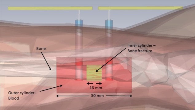
For comparison, they simulated bone fractures in a voxel model of a 26-year-old female, with a fracture represented by two coaxial cylinders. The inner cylinder was injected with blood-simulating tissue to represent the dielectric properties of various stages of healing. The outer cylinder surrounding the “bone” was given the dielectric properties of blood. They simulated the maximum amount of haematoma at the initial fracture state, and gradually reduced it until it was removed entirely.
Finally, the team conducted an ex-vivo measurement using a lamb femur bone. Extensive testing confirmed that, in all cases, the power transmitted from one monopole antenna to the other through the fracture decreased significantly as the volume of blood increased.
The authors note that the pattern of change of S21 could be observed in much of the frequency range of 1 to 4 GHz. Monopole lengths could easily be adapted to fit the diameter and depth of any human bone likely to have external fixators. Because no two fractures are identical, considerable variation in S21 magnitude is anticipated. However, if a healing time could be estimated, the rate of change of S21 would expect to slow over this period. This information could indicate that bone healing had stalled.
While initial results are highly promising, there is still a long way to go before this technique can be used in clinical trials. Further activities will include comparison with other, more traditional techniques, moving slowly away from the workbench into clinical assessment.


