A microscope that can track the real-time motions of individual blood cells throughout the entire brains of living mice has been created by Qionghai Dai at Tsinghua University in China and colleagues.
Their real-time, ultra-large-scale, high-resolution (RUSH) system combines a large field of view, with high spatial and temporal resolution. The microscope could be used for medical and biological imaging, allowing researchers to observe the complex, ever-changing characteristics of active living systems. The ability to acquire such videos could prove to be incredibly useful in medical imaging, since the complex dynamics of individual parts in biological systems are almost impossible to measure using conventional techniques.
A conventional optical microscope can either have a large magnification (high spatial resolution) or a large field of view (FOV) but not both. In 2016, a team of researchers UK unveiled a “mesolens” microscope, which offered both features. Their imaging platform stitches together images taken by 15 extremely precise optical elements. This minimizes the aberrations plaguing previous designs of confocal microscopes and allowed the system to image sub-cellular details across an entire mouse embryo.
Vast amounts of data
Now, Dai’s team have gone one step further with their RUSH system, which captures large FOV, high resolution videos of time-varying biological dynamics. In doing so they have overcome an important challenge: at large FOVs and high resolutions, a video microscope will generate vast amounts of data that must be handled in an efficient way.
‘Radical’ new microscope lens combines high resolution with large field of view
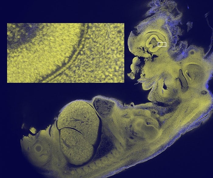
Dai and colleagues dealt with this data deluge by designing the RUSH objective lens to imitate the compound eyes of insects. This was done by arranging the microscope’s optical elements in a concave mosaic. When taking images, the sample plane was magnified onto the curved surface, before the images taken by each element were conveyed to an array of planar sensors, where they were stitched together to rebuild the original image. This setup reduced aberrations even further compared with previous platforms, allowing the researchers to take videos at centimetre-scale FOVs, micron resolution, and with a data throughput as high as 5.1 gigapixels per second.
With their setup, Dai’s team used fluorescent dyes to track the motions of individual white blood cells throughout the entire brains of active living mice, at speeds of up to 30 frames per second. They say that with further improvements, their updated RUSH platform could allow for the functional imaging of activity in intercellular networks, as well as the structural imaging of cellular dynamics in cultured cells, human brain tissues and awake, behaving animals. This would ultimately open up new opportunities for improved diagnoses and treatments, including high-speed screening for pathogens, as well as real-time imaging of tumour metastasis and neuron activity.
The new microscope is described in Nature Photonics.