Respiration-correlated CT imaging, or 4D CT, is an essential part of radiotherapy planning for thoracic and abdominal tumours. Clinical 4D CT images are often affected by artefacts, however, mainly due to irregular breathing patterns during data acquisition. A research team has now successfully validated a prototype implementation of an intelligent 4D CT (i4DCT) scanning protocol that produces fewer motion artefacts, demonstrating that breathing signal-guided 4D CT is feasible for clinical applications.
The researchers, at the University Medical Center Hamburg-Eppendorf (UKE) in Germany and Siemens Healthineers, utilized online breathing curve analysis and respiratory signal-guided 4D CT protocols to develop the i4DCT concept. In a feasibility study, they compared images of a motion phantom recorded using routine spiral 4D CT and i4DCT (Med. Phys. 10.1.1002/mp.14106).
4D CT images are used for dose calculation and optimization, to define motion-adapted safety margins, and to perform 4D dose reconstruction and quality assurance after radiation treatment. When images are degraded by artefacts, it may be necessary for a patient to have an additional CT scan, exposing them to a second high radiation dose and potentially delaying their radiotherapy.
The i4DCT concept enables automated selection of CT beam-on/beam-off periods, by adapting data acquisition to a patient’s individual breathing pattern, instead of the patient having to adapt to the scanner. The i4DCT workflow consists of an initial learning period to establish a reference patient-specific breathing cycle representation, followed by online breathing signal-guided sequence mode scanning.
The process involves switching on the CT beam at a breathing state just before the patient’s typical end-inspiration state, continuously acquiring breathing signal and projection data, simultaneously analysing the breathing signal in terms of projection data coverage, and switching off the beam if predefined coverage conditions are fulfilled. The i4DCT monitors the patient’s breathing in real time, starting and stopping the sequence scanning based on online analysis of the acquired breathing curve information.
After a beam-off event, which corresponds to a breathing state close to, but after, end-inspiration, the scanner couch is moved to the next position and the process repeated until the desired scanning range is covered. The projection data are then used for retrospective image reconstruction.
Protocol validation
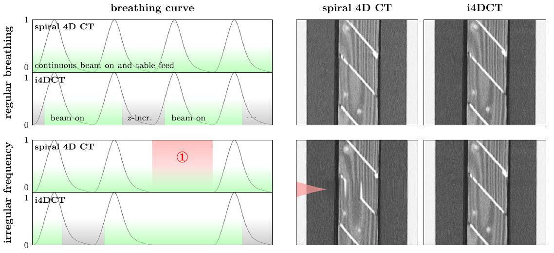
For the study, the researchers implemented the i4DCT core workflow on a Siemens SOMATOM go platform, and imaged a motion phantom containing a customized wooden insert with oblique aluminium plates. They examined four programmed motion curves: regular breathing; breathing pause/irregular breathing frequency; irregular breathing amplitude; and a mixture of breathing pause, and frequency and amplitude irregularity.

Principal investigators René Werner and Christian Hofmann reported that in a regular breathing scenario, both routine spiral 4D CT and i4DCT generated similar images. Spiral 4D CT images acquired during the breathing pause/irregular breathing frequency scenario contained interpolation artefacts that distorted the appearance of the central aluminium plate. However, the i4DCT acquired data did not have these artefacts. For the other two motion scenarios, i4DCT-acquired data also outperformed spiral 4D CT data, producing images of better quality with fewer artefacts.
The researchers point out that i4DCT will not generate artefact-free images for every patient and every breathing pattern. Because it relies on external breathing signals, a robust correlation of internal and external motion data is needed to produce artefact-free images.
The i4DCT scanning time was also longer: by 38%, 72%, 82% and 100%, respectively, for the four motion scenarios compared with conventional 4D CT. With the exception of the regular breathing scenario, beam-on time also was longer, but only by 13%, 20% and 25% compared with conventional 4D CT.
“The measurement results and acquired images support the conclusions of the [previously published] in silico studies and illustrate the considerable reduction in 4D CT image artefacts by real-world application of i4DCT and comparison to routine spiral 4D CT,” the authors write.
“In line with this statement, the i4DCT functionality has been integrated into Siemens go.Open Pro scanners and is called Direct i4D technology,” Werner tells Physics World. “Currently, the collaboration partners are working on a comprehensive phantom-based study to double check the Direct i4D performance for patient breathing patterns under real-world conditions. Moreover, together with other radiotherapy affiliations, the first 4D CT images of lung and liver patients have been measured and are under evaluation.”