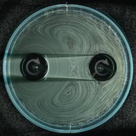
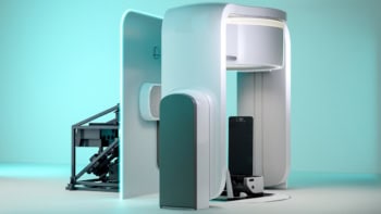
A "lensless" x-ray microscope that can take pictures of biological samples in their natural environment has been developed in the UK. Physicists used several overlapping diffraction patterns to create a wide field of view of samples, which they claim provides virtually instantaneous images with a resolution limited only by the x-rays' wavelength. The principle could also be implemented in a new type of large-scale 3D imaging akin to medical CT scans (Phys. Rev. Lett. 98 034801).
Lenses for x-rays are notoriously tricky to manufacture because they rely on nanometre-scale features. For this reason, physicists developing x-ray microscopes have been keen to adopt “lensless” designs that measure diffraction when x-rays are passed through samples. But while this principle has worked well for periodic structures such as crystals, methods for non-periodic structures such as biological materials have been ineffective.
In existing lensless x-ray microscopes, data from just a single diffraction measurement are taken, which must then be subjected to an algorithm that gradually hones in on a “solution” or image after many thousands of steps. At the University of Sheffield, however, John Rodenburg and colleagues have used a sophisticated technique dating from 1969 called “ptychography” that melds many overlapping diffraction measurements together, meaning only a few steps are required before a detailed image is resolved.
A crude analogy for this, according to Rodenburg, would be to imagine clapping you hands while blindfolded in a mountain range. If you were to remain stationary, it would be very difficult to work out the positions of the mountains by hearing the echoes alone. But if you were to steadily move around, it would be much easier to build up a rough picture of where they all are.
The key advantage of the technique when applied to x-ray microscopy is that the resultant image has a very wide field of view, so the portion of a sample under inspection can be immediately recognized. Not only does this enable biological samples to be examined in their natural environment, but the wavelength-scale detail is such that segments of the image can be enlarged to be viewed more clearly.
Rodenburg said that a large-scale version of the device could be used to take 3D images like modern CT scans in hospitals. However, he added that the lensless design principle could be extended to other parts of the spectrum that are unable to be focused optically, such as ultraviolet or terahertz wavelengths. “We can get as good an image as the best optical microscope in the world,” he said.


