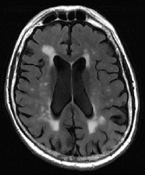
Every year, there are 10 million new cases of dementia, according to the World Health Organization. Despite this, our understanding of this condition is limited and our ability to identify its signs in the brains of patients is crude. New research from NYU Langone Health may improve our ability to identify early signs of dementia and cognitive decline from a patient’s brain scans.
How to spot the difference
Diagnosing dementia is particularly challenging for doctors, with no test able to give a straightforward “yes or no” answer. What’s more, even the tools we do have are limited. An MRI scan allows us to see into the brain and observe bright spots, called white matter hyperintensities.

Having more of these spots, especially in the centre of the brain, is linked to dementia and other conditions that cause damage to the brain. Right now, though, identifying these spots and their extent is just a matter of having a “trained eye”, according to researchers behind a new study in the journal Academic Radiology.
The team, led by Jingyun Chen from the department of neurology and Yulin Ge from the department of radiology, aimed to produce a more objective way to characterize these bright spots on brain scans. To achieve this, the researchers developed a new algorithm that is able to identify two different types of bright spots more consistently than alternative approaches.
This information could be used to distinguish between brain scans from healthy patients, patients with cognitive decline and patients diagnosed with Alzheimer’s disease more accurately than other methods. In fact, in tests on 72 MRI scans from 60 subjects, seven out of 10 of the algorithm’s predictions matched the patient’s clinical diagnosis.
Putting brain scans to the test
The method works by finding the precise position and volumes of all white matter lesions present in the scans. These are then analysed and classified based on the distance to both sides of the brain, to characterize which are the more concerning spots close to the centre of the brain and how large they are.
“Our new calculator for properly sizing white matter hyperintensities, which we call bilateral distancing, offers radiologists and other clinicians an additional standardized test for assessing these lesions in the brain, well before severe dementia or stroke damage,” says Ge.
Whilst the researchers are clear that their tool cannot be used alone to diagnose dementia, “amounts of white matter lesions above the normal range should serve as an early warning sign for patients and physicians,” according to Chen.
Having started with a limited series of scans, the team aims to analyse hundreds more to hone their method and has made the code freely available to other researchers and clinicians. As systematic methods for analysing these scans continue to improve, doctors will soon hopefully be able to use more than just their eyes to routinely track down key signs of brain disease.


