
Researchers at Pohang University of Science and Technology (POSTECH) and Seoul National University (SNU) have fabricated a biomimetic airway-on-a-chip using a 3D printing technique that exploits bioinks laden with cells taken from human trachea. The device, which contains a network of blood vessels connecting with epithelial cells, could be used as a model to study respiratory diseases, such as asthma, rhinosinusitis, and chronic lung disease.
Rising air pollution in many counties is both increasing the number of people who suffer from respiratory diseases, and making their symptoms more severe. Researchers have therefore attempted to create life-like models that could be used to characterize these inflammatory diseases.
In this new research, a team led by Dong-Woo Cho made a biomimetic airway structure by 3D printing the various different types of cells that are found in natural mucous membranes. The bioink was composed of a decellularized extracellular matrix (dECM) isolated from pig trachea, which was laden with mucosal isolated from human trachea (Biofabrication 11 015002).
“We reproduced an in-vivo-like 3D vascular network by assembling endothelial cells and fibroblasts using the dECM bioink in a one-step printing process,” explains lead author of the study, Ju Young Park. “The structure we produced has the same physiological functions as the biological airway epithelium and so can be used to model diseases like asthma. The presence of blood vessels, for example, leads to an excessive production of proinflammatory cytokines in our airway model. This process (also known as the ‘cytokine storm’) occurs during asthmatic airway inflammation and allergen-induced asthma exacerbation in the physiological context.”
The researchers also confirmed that their model epithelium airway becomes sensitized by house dust mites, which are well-known respiratory allergens. These allergens stimulate the expression of an adhesion molecule on the vascular endothelium that can then recruit immune cells into the inflamed tissue. “Our results show that pathological interactions between the airway endothelium and the vascular network in the airway are reproducible in our airway-on-a-chip model, and that the exacerbation of inflammatory responses by the vascular network in vivo is also reproducible in vitro.”
Mimicking complex 3D structures
The Korean team printed its structures using an in-house 3D cell printer equipped with six dispensing heads. “Two of the printing heads were connected to a pneumatic pressure-based system that dispenses a synthetic polymer to fabricate the supporting framework for the airway,” explains Park. “The other four printing heads operate on a three-axis motorized stage and we control their movement using computer programs.”
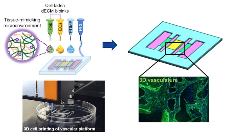
Park explains that the epithelium in the human airway mainly contains ciliated goblet and basal cells in contact with basement membrane. Lamina propria, which contain blood vessels and stromal fibroblasts, lie under this membrane. “To mimic this complex 2D/3D structure and the cellular composition of the airway mucosa, we assembled a 2D airway epithelium on a 3D vascular platform,” she says. “We reconstructed the natural 3D vascular network by 3D cell printing the dECM bioink containing endothelial cells and fibroblasts. The dECM bioink in fact provides the cell with an in-vivo-like niche of native tissue that induces tissue-specific differentiation and function.”
According to Park, the cell-printing technique is much faster than traditional fabrication techniques, and is also better at replicating the fine cellular arrangement and complex 3D microstructure found in natural tissue. “Our 3D cell-printing system allows us to easily fabricate airway prototypes in high throughput and also allows us to directly place various types of cell at specific locations on the airway structure to mimic how cells arrange themselves in native tissue,” she explains. “The technique could be used to design many types of chip and even print organ models other than the airway.”
Towards multiple organs-on-a-chip
The interaction between the epithelium and the blood vessels in the mucous membrane is known to be important for the health of the tissue and to protect against allergens or toxins. “Our new model could be used to study these interactions and better understand the role they play in human respiratory diseases,” says Park. “The 3D cell printed airway-on-a-chip could therefore be used as a powerful complement to animal models for analysing pathophysiology and testing the efficiency of drugs in the preclinical phase.” This technology is currently under development for commercialization by T&R Biofab, a Korean company that makes biomedical products using 3D cell printing technology.
The researchers, reporting their work in the journal Biofabrication, say that they would now like to 3D cell print different organs-on-a-chip and even multiple organs-on-a-chip. These structures would integrate a number of interacting tissues and organs, and could ultimately replace animal models for studying human pathophysiology and evaluating systemic drug effects.
- Read our special collection “Frontiers in biofabrication” to learn more about the latest advances in tissue engineering. This article is one of a series of reports highlighting high-impact research published in Biofabrication.


