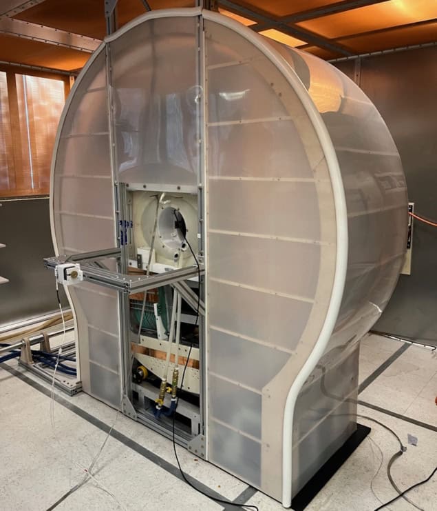
Magnetic particle imaging (MPI) is an emerging medical imaging modality with the potential for high sensitivity and spatial resolution. Since its introduction back in 2005, researchers have built numerous preclinical MPI systems for small-animal studies. But human-scale MPI remains an unmet challenge. Now, a team headed up at the Athinoula A Martinos Center for Biomedical Imaging has built a proof-of-concept human brain-scale MPI system and demonstrated its potential for functional neuroimaging.
MPI works by visualizing injected superparamagnetic iron oxide nanoparticles (SPIONs). SPIONs exhibit a nonlinear response to an applied magnetic field: at low fields they respond roughly linearly, but at larger field strengths, particle response saturates. MPI exploits this behaviour by creating a magnetic field gradient across the imaging space with a field-free line (FFL) in the centre. Signals are only generated by the unsaturated SPIONs inside the FFL, which can be scanned through the imaging space to map SPION distribution.
First author Eli Mattingly and colleagues propose that MPI could be of particular interest for imaging the dynamics of blood volume in the brain, as it can measure the local distribution of nanoparticles in blood without an interfering background signal.
“In the brain, the tracer stays in the blood so we get an image of blood volume distribution,” Mattingly explains. “This is an important physiological parameter to map since blood is so vital for supporting metabolism. In fact, when a brain area is used by a mental task, the local blood volume swells about 20% in response, allowing us to map functional brain activity by dynamically imaging cerebral blood volume.”
Rescaling the scanner
The researchers began by defining the parameters required to build a human brain-scale MPI system. Such a device should be able to image the head with 6 mm spatial resolution (as used in many MRI-based functional neuroimaging studies) and 5 s temporal resolution for at least 30 min. To achieve this, they rescaled their existing rodent-sized imager.

The resulting scanner uses two opposed permanent magnets to generate the FFL and high-power electromagnet shift coils, comprising inner and outer coils on each side of the head, to sweep the FFL across the head. The magnets create a gradient of 1.13 T/m, sufficient to achieve 5–6 mm resolution with high-performance SPIONs. To create 2D images, a mechanical gantry rotates the magnets and shift coils at 6 RPM, enabling imaging every 5 s.
The MPI system also incorporates a water-cooled 26.3 kHz drive coil, which produces the oscillating magnetic field (of up to 7 mTpeak) needed to drive the SPIONs in and out of saturation. A gradiometer-based receive coil fits over the head to record the SPION response.
Mattingly notes that this rescaling was far from straightforward as many parameters scale with the volume of the imaging bore. “With a bore about five times larger, the volume is about 125 times larger,” he says. “This means the power electronics require one to two orders of magnitude more power than rat-sized MPI systems, and the receive coils are simultaneously less sensitive as they become larger.”
Performance assessment
The researchers tested the scanner performance using a series of phantoms. They first evaluated spatial resolution by imaging 2.5 mm-diameter capillary tubes filled with Synomag SPIONs and spaced by between 5 and 9 mm. They reconstructed images using an inverse Radon reconstruction algorithm and a forward-model iterative reconstruction.
The system demonstrated a spatial resolution of about 7 mm with inverse Radon reconstruction, increasing to 5 mm with iterative reconstruction. The team notes that this resolution should be sufficient to observe changes in cerebral blood volume associated with brain function and following brain injuries.
To determine the practical detection limit, the researchers imaged Synomag samples with concentrations from 6 mgFe/ml to 15.6 µgFe/ml, observing a limit of about 1 µgFe. Based on this result, they predict that MPI should show grey matter with a signal-to-noise ratio (SNR) of roughly five and large blood vessels with an SNR of about 100 in a 5 s image. They also expect to detect changes during brain activation with a contrast-to-noise ratio of above one.
Next, they quantified the scanner’s imaging field-of-view using a G-shaped phantom filled with Synomag at roughly the concentration of blood. The field-of-view was 181 mm in diameter – sufficient to encompass most human brains. Finally, the team monitored the drive current stability over 35 min of continuous imaging. At a drive field of 4.6 mTpeak, the current deviated less than 2%. As this drift was smooth and slow, it should be straightforward to separate it from the larger signal changes expected from brain activation.
Multimodal MRI reveals brain areas that can still ‘see’ after a stroke
The researchers conclude that their scanner – the first human head-sized, mechanically rotating, FFL-based MPI – delivers a suitable spatial resolution, temporal resolution and sensitivity for functional human neuroimaging. And they continue to improve the device. “Currently, the group is developing hardware to enable studies such as application-specific receive coils to prepare for in vivo experiments,” says Mattingly.
At present, the scanner’s sensitivity is limited by background noise from the amplifiers. Mitigating such noise could increase sensitivity 20-fold, the team predicts, potentially providing an order of magnitude improvement over other human neuroimaging methods and enabling visualization of haemodynamic changes following brain activity.
The MPI system is described in Physics in Medicine & Biology.


