Magnetic resonance imaging (MRI) is one of the most common techniques used to image internal body parts of animals and humans, in particular for cancer diagnosis. At a resolution of some few micrometres, conventional MRI instruments need relatively large samples to provide good images. In recent times, various research groups have sought to obtain resolutions down to atomic sizes. In the most recent breakthrough, a group of scientists from the US and Korea, led by Andreas Heinrich at the Ewha Womans University and the associated IBS Center for Quantum Nanoscience in Korea and Christopher Lutz at IBM Almaden in the US, has imaged single atoms on a surface. This boosts the potential of MRI for studying biomolecules with unprecedented resolution, as well as investigating the spin structure of atoms, molecules, solids, quantum systems and spin networks.
One of the quantum properties used to characterize protons, neutrons and electrons is their spin, which is related to the presence or absence of magnetic properties in a material. Conventional MRI is based on nuclear magnetic resonance, which measures the spin properties of atomic nuclei. To improve the resolution of the images Heinrich and Lutz and co-workers exploited scanning tunnelling microscopy (STM) – a technique that can resolve atoms by monitoring the tunnelling current between a tip and surface as it scans across a sample. STM resolution greatly exceeds what has been achieved by other alternative approaches to image spin properties but previous attempts to measure magnetic interactions with STM have been hampered by thermal motion. To image spin properties with atomic resolution the researchers use STM to detect changes in the electron spin resonance at cryogenic temperatures.
Combined techniques
They first adsorb single atoms onto a two-atom-thick layer of magnesium oxide and create a spin cluster on the STM tip by adding iron atoms. The conducting tip then provides the magnetic field gradient, electric read-out and the driving field required for the measurements. Applying a radio frequency (RF) voltage induces a transition between different spin states, so that at the atomic resonance frequency the tunnelling current peaks in the tip brought close to the surface. The magnetic field from the tip splits the spin states into additional energy levels so that the resonant frequency varies across the atom. Consequently scanning the atom scanned with the tip at a constant frequency maps out these spatially varying resonance patterns providing an atomic resolution spin image.
How does MRI work?

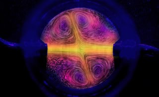
The scientists mapped out the three-dimensional (3D) magnetic interaction potential between spins of the magnetic tip and the surface atom. In addition, they show how various tip configurations and atomic species revealed details of the magnetic properties of both tip and surface atoms.
This work is published in Nature Physics.