
A new scanning technique that uses hyperpolarized carbon-13 MRI to monitor the metabolism of different types of breast cancer can identify how rapidly a tumour is growing. The technique, developed by researchers at Cancer Research UK Cambridge Institute and the University of Cambridge, could help doctors prescribe the best course of treatment for a patient and follow how they respond.
Breast cancer accounts for roughly a quarter of all cancer cases worldwide and is the leading cause of cancer death in women. There are many subtypes of breast cancer, with some being more aggressive than others. The new technique is the first to be able to detect differences in a tumour’s size, type and grade (a measure of how fast it is growing), while also yielding information on the variations in metabolism between different regions within the tumour.
Measuring pyruvate metabolism
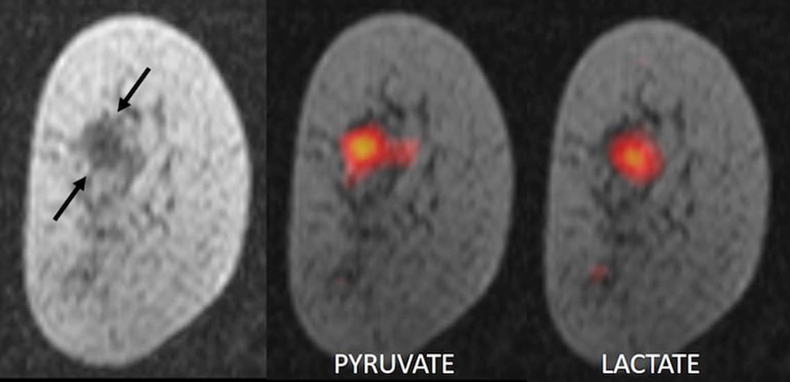
The technique works by measuring how fast a tumour metabolizes a naturally-occurring sugar-like molecule called pyruvate. In their experiments, the researchers used pyruvate that they had labelled with carbon-13, a heavier isotope of carbon. They then hyperpolarized, or magnetized, this carbon-13 pyruvate by cooling it to -272°C and exposing it to an extremely strong magnetic field and microwave radiation in a special machine called SPINlab. The frozen pyruvate sample was then thawed, dissolved into a solution and injected into the patient, who immediately underwent an MRI scan.
Tumours make use of large amounts of sugar and take up more pyruvate than normal tissue, explains team member Ramona Woitek. Inside the tumour, pyruvate is converted into lactate as part of a natural metabolic process. Since magnetizing the carbon-13 pyruvate molecules increases the strength of the MRI signal by 10 000 times, the researchers can monitor this process and visualize it dynamically in MRI scans.
The rate of pyruvate metabolism – and the amount of lactate produced – varies not only between different tumours, but also between different regions of the same tumour. By monitoring this conversion in real-time, the researchers say they are able to determine the type of cancer being imaged. They can also determine how aggressive a tumour is, as faster growing tumours convert pyruvate more rapidly than less aggressive ones.
Monitoring chemotherapy
The technique will be useful for monitoring patients undergoing chemotherapy, says Woitek. It will allow determination of how efficient a given treatment is by imaging a tumour before and after the therapy, and repeatedly during the course of treatment. “Identifying patients that do not respond to a treatment will thus allow us to change the therapeutic strategy early on,” she says. “Conversely, we may even be able to reduce a treatment dose if a patient is responding well, thus sparing them unnecessary side effects.”
“Researchers are beginning to understand that the many different types of breast cancer respond differently to different treatments,” Woitek tells Physics World. “The new hyperpolarized carbon-13 MRI approach could allow us to identify the optimal treatment for each individual patient.”
The researchers, who report their work in PNAS, have tested their technique on seven patients, all with different types and grades of cancer. They say they now hope to study larger groups of patients.


