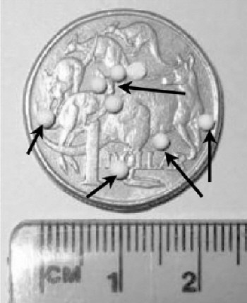
Researchers in Australia have developed a new technique to make small sources of radioactivity for applications in nuclear medicine. Dale Bailey and colleagues at the Royal North Shore Hospital in Sydney made their compact sources by soaking small beads in a solution of radioactive liquid for a few minutes. The beads, which are removed with tweezers, could be used to measure the improved spatial resolution of various new medical imaging techniques (D Bailey et al. 2004 Phys. Med. Biol. 49 N21)

Radioactive tracers are routinely used in biomedical imaging and offer a non-invasive way to look inside the body. Positron emission tomography (PET), for instance, can now achieve resolutions of between 4 and 8 millimetres, but testing this improved resolution involves, among other things, making the sources of the radioactivity compact. Researchers have tried to make such sources by irradiating wires or small particles, but this has proved difficult to do routinely.
Bailey and co-workers soaked commercially available beads of an aluminium-silicon compound in a solution of radioactive technetium-99. The beads had an average diameter of about 2.1 millimetres and contained millions of tiny pores – which is why they are sometimes called molecular sieves. The sieves absorbed and trapped molecules from the solution in their pores, and a typical bead displayed a radioactivity of between 3 to 6 megabecquerels after being soaked for two minutes.
“I have presented this technique at a couple of conferences and am amazed at how much interest it sparks,” Bailey told PhysicsWeb. “Making such small compact sources has clearly been elusive and the fact that this method is so easy – and inexpensive – means that anybody could use it,” he said.
The Australian researchers say that smaller-sized sieves produce higher levels of radioactivity and could therefore be used to test even sharper images. This could important for the new generation of animal scanners that have a spatial resolution of about 1 to 2 millimetres. Moreover, the beads can also absorb fluorine-18, a tracer widely used in PET. The team now plans to investigate whether these sources are visible with other scanning techniques, such as X-ray or magnetic resonance imaging (MRI).


