From tumour-killing quantum dots to proton therapy firsts, this year has seen the traditional plethora of exciting advances in physics-based therapeutic and diagnostic imaging techniques, plus all manner of innovative bio-devices and biotechnologies for improving healthcare. Indeed, the Physics World Top 10 Breakthroughs for 2024 included a computational model designed to improve radiotherapy outcomes for patients with lung cancer by modelling the interaction of radiation with lung cells, as well as a method to make the skin of live mice temporarily transparent to enable optical imaging studies. Here are just a few more of the research highlights that caught our eye.
Marvellous MRI machines
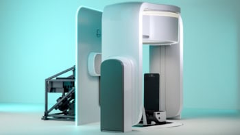
This year we reported on some important developments in the field of magnetic resonance imaging (MRI) technology, not least of which was the introduction of a 0.05 T whole-body MRI scanner that can produce diagnostic quality images. The ultralow-field scanner, invented at the University of Hong Kong’s BISP Lab, operates from a standard wall power outlet and does not require shielding cages. The simplified design makes it easier to operate and significantly lower in cost than current clinical MRI systems. As such, the BISP Lab researchers hope that their scanner could help close the global gap in MRI availability.
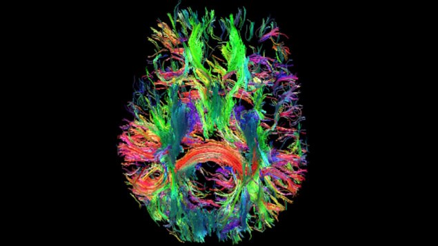
Moving from ultralow- to ultrahigh-field instrumentation, a team headed up by David Feinberg at UC Berkeley created an ultrahigh-resolution 7 T MRI scanner for imaging the human brain. The system can generate functional brain images with 10 times better spatial resolution than current 7 T scanners, revealing features as small as 0.35 mm, as well as offering higher spatial resolution in diffusion, physiological and structural MR imaging. The researchers plan to use their new NexGen 7 T scanner to study underlying changes in brain circuitry in degenerative diseases, schizophrenia and disorders such as autism.
Meanwhile, researchers at Massachusetts Institute of Technology and Harvard University developed a portable magnetic resonance-based sensor for imaging at the bedside. The low-field single-sided MR sensor is designed for point-of-care evaluation of skeletal muscle tissue, removing the need to transport patients to a centralized MRI facility. The portable sensor, which weighs just 11 kg, uses a permanent magnet array and surface RF coil to provide low operational power and minimal shielding requirements.
Proton therapy progress
Alongside advances in diagnostic imaging, 2024 also saw a couple of firsts in the field of proton therapy. At the start of the year, OncoRay – the National Center for Radiation Research in Oncology in Dresden – launched the world’s first whole-body MRI-guided proton therapy system. The prototype device combines a horizontal proton beamline with a whole-body MRI scanner that rotates around the patient, a geometry that enables treatments both with patients lying down or in an upright position. Ultimately, the system could enable real-time MRI monitoring of patients during cancer treatments and significantly improve the targeting accuracy of proton therapy.

Also aiming to enhance proton therapy outcomes, a team at the PSI Center for Proton Therapy performed the first clinical implementation of an online daily adaptive proton therapy (DAPT) workflow. Online plan adaptation, where the patient remains on the couch throughout the replanning process, could help address uncertainties arising from anatomical changes during treatments. In five adults with tumours in rigid body regions treated using DAPT, the daily adapted plans provided target coverage to within 1.1% of the planned dose and, in over 90% of treatments, improved dose metrics to the targets and/or organs-at-risk. Importantly, the adaptive approach took just a few minutes longer than a non-adaptive treatment, remaining within the 30-min time slot allocated for a proton therapy session.
Bots and dots
Last but certainly not least, this year saw several research teams demonstrate the use of tiny devices for cancer treatment. In a study conducted at the Institute for Bioengineering of Catalonia, for instance, researchers used self-propelling nanoparticles containing radioactive iodine to shrink bladder tumours.

Upon injection into the body, these “nanobots” search for and accumulate inside cancerous tissue, delivering radionuclide therapy directly to the target. Mice receiving a single dose of the nanobots experienced a 90% reduction in the size of bladder tumours compared with untreated animals.
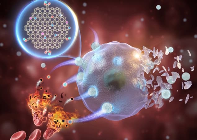
At the Chinese Academy of Sciences’ Hefei Institutes of Physical Science, a team pioneered the use of metal-free graphene quantum dots for chemodynamic therapy. Studies in cancer cells and tumour-bearing mice showed that the quantum dots caused cell death and inhibition of tumour growth, respectively, with no off-target toxicity in the animals.
Finally, scientists at Huazhong University of Science and Technology developed novel magnetic coiling “microfibrebots” and used them to stem arterial bleeding in a rabbit – paving the way for a range of controllable and less invasive treatments for aneurysms and brain tumours.


