Physicists in Spain have showed that metamaterials — artificial materials with exotic electromagnetic properties — can dramatically increase the sensitivity of magnetic resonance imaging (MRI). As well as enabling the technique to probe into deeper tissue, the researchers say that metamaterials could also enhance image quality and cut the amount of time taken to acquire images.
MRI has become a standard technique in medical diagnostics and chemical analysis over the past three decades. It involves placing a substance in a fixed magnetic field while exposing it to radio waves, which become absorbed by the substance’s atomic nuclei. By mapping the variations in phase and frequency of the absorbed radio waves with a receiving coil, a scientist can create an image of the substance’s internal structure.
Ricardo Marques and colleagues from the University of Seville in Spain have found that metamaterials can extend the depth to which a receiving coil is sensitive. They are not the first researchers to discover that certain materials can increase the capabilities of MRI — in 2001 a team including John Pendry of Imperial College, London, found that a material with a very high refractive index can be placed on top of the substance to act as a “flux guide” so that the receiving coil can be positioned farther away. By contrast, Marquez’s team has used a metamaterial with a negative refractive index to shift the magnetic field deep inside a patient to the outside (arXiv:0810.1689).
Both knees
In their experiment, Marquez and colleagues first performed a standard MRI scan of a male patient with the receiving coil held beside his knees. They found that they could image a cross section roughly as deep as one knee, but not the other.
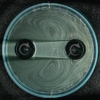
Next they tried placing a slab of negative-index metamaterial between his knees. The metamaterial, which was 27 cm square and 3 cm thick, consisted of a 3D cubic array of copper rings that were each loaded with a capacitor. As the researchers performed another MRI scan, they found that the resultant cross-section image extended through both knees (see figure).
The researchers say that the ability to probe significantly deeper could reduce the time it takes to scan a large volume, and could also lead to improved image quality. In fact, they have already patented their technique and are working with a Spanish company called PET Cartuja to develop it further.
However, Marques told physicsworld.com that his team still needs to make it more adaptable. In normal MRI scans the receiver coil has to be tuned to the type of substance being probed — but in the Spanish team’s technique, the metamaterial must also be tuned.


