Researchers in Switzerland have obtained the first direct images of magnetization being transported through a material by the diffusion of nuclear spins. The work was done using a special magnetic resonance imaging (MRI) microscope developed by the team, who claim that their achievement will lead to a better understanding of nuclear spin diffusion – which plays an important role in nuclear magnetic resonance (NMR) studies of large molecules such as proteins and polymers.
Nuclear magnetic resonance works by applying a strong magnetic field to a material, which lines up its nuclear spins. The spins are then knocked out of alignment by applying radio-frequency signals – a process that can deliver a wealth of information about the chemical and structural properties of the material.
Flip-flop transitions
Spin diffusion is a process by which nuclear spins transport magnetization from one region of a solid to another and it plays an important role in how a material responds to NMR. Diffusion occurs through a series of “flip-flop” transitions involving pairs of neighbouring spins — a pair with spins pointing “up” and “down” respectively is transformed into a pair with spins pointing “down” and “up”, for example. These flip-flops tend to even-out imbalances between the numbers of up and down spins in a region of a solid by diffusing excess spins away.
Spin diffusion was discovered nearly 60 years ago and its effects have been used to determine molecular distances in NMR studies of proteins. However, physicists had been unable to actually watch the process occur in space and time. This is because diffusion occurs over distances of only a few hundred nanometres – and until very recently, it was impossible to obtain MRI images at this spatial resolution.
The magnetic resonance force microscope allows us to image the magnetization distribution at length scales small enough to directly visualize the spin-diffusion process
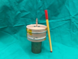
Now, Kai Eberhardt and Beat Meier at ETH Zurich, along with their colleagues at EPFL in Lausanne have used a relatively new technique called magnetic resonance force microscopy (MRFM) to see spin diffusion for the first time (Phys. Rev. Lett. 99 227603 ). The team mounted their sample – a calcium fluoride crystal 25 micrometres across – on a tiny cantilever. The sample and cantilever are placed in a 6 Tesla magnetic field near to an iron tip, which creates a magnetic field gradient in the region of the sample. A coil is also placed near to the sample, which broadcasts a radio signal that causes the cantilever to vibrate.
The magnetic force on the cantilever is determined by how many spin up and spin down nuclei are in the sample. By carefully monitoring the motion of the cantilever as it vibrates back and forth through the magnetic field gradient, changes in this force and therefore changes in the directions of spins can be determined. In this way, the team are able to measure the magnetization of the fluorine nuclei in the sample at nanometre distances.
To do this, Meier and his colleagues first magnetized the sample and then allowed spin diffusion to evolve and propagate through the system. MFRM was then used to obtain 1D images of the changing magnetization at regular time intervals. From these images, the team was able to calculate the spin diffusion rate for calcium fluoride – which was in agreement with values previously measured using different techniques.
Better understanding
“The magnetic resonance force microscope allows us to image the magnetization distribution at length scales small enough to directly visualize the spin-diffusion process,” Meier told physicsworld.com. “This will let us study this fundamental process directly and understand it better.”
A better understanding of spin diffusion could lead to improvements in the way that NMR is used to study the structure of proteins and polymers. The ability to measure the movement of spin on the nanometre scale could also help in the development of tiny “spintronic” devices that use both the spin and charge of the electron to store and process information. Eberhardt told physicsworld.com that the team are now working on a way to obtain 3D images of spin diffusion in materials – something that would allow the technique to be used to study spintronic systems.