Treatment of epidural tumours located in the spinal canal can be highly challenging, due to the risk of causing irreversible damage. To address this challenge, a medical team at Brigham and Women’s Hospital and Dana Farber Cancer Institute have performed a study showing that MRI-guided cryoablation is feasible for treating such epidural tumours. After cryoablation, both patients in the study experienced successful decompression of the tumour away from the spinal cord, regrowth of previously eroded bone around the spinal canal, and reduction or elimination of the risk of becoming paralysed by the cancer (Am. J. Roentgenol. 10.2214/AJR.18.19951).
When a tumour compresses the spinal cord, previous options treatment included spinal decompression, a surgical procedure to relieve pressure, spinal decompression and radiotherapy, stereotactic radiosurgery, or laser interstitial thermal therapy. Each of these treatments, however, is associated with serious risks. Some patients are unsuitable candidates for surgery, and some who undergo it may not recover rapidly, delaying radiotherapy. Radiation dose delivery may be limited by the tumour’s proximity to the spinal cord, therefore reducing the effectiveness of radiotherapy. And ablative techniques such as laser interstitial thermal therapy may cause permanent neural burn injury.
“The main advantage of cryoablation over heat-based ablation modalities such as radiofrequency ablation is visualization of a distinct edge of ablation, which can be monitored as a black ovoid region on standard MRI,” the authors state. “Another advantage of cryoablation … is the reduced risk of injuring adjacent structures, such as carotid artery walls, leading to dissection and blowout, or central nervous structures, which have no ability to regenerate.”
Cryoablation also enables a patient to regrow previously eroded bone and regenerate cortex and marrow architecture, which does not happen after surgical resection or heat-based ablative treatments.
MRI provides excellent visualization and nearly real-time imaging of the tumour, the ablation zone and involved neural structures. It is also better than CT at visualizing the cryoablation zone and the adjacent spinal cord as, with CT, the high radiodensity of bone can obscure the low-radiodensity ice ball. Unlike CT, MRI can also be used to accurately image epidural tumours invading or compressing central nervous system structures.
The MRI-guided cryoablation process
Lead author Thomas Lee, a neuroradiologist at Brigham and Women’s Hospital, told Physics World that the hospital’s MRI-guided operating suite was made possible through the vision of the late neuroradiologist Ferenc Joelsz, known for his breakthrough contributions in image-guided therapy.
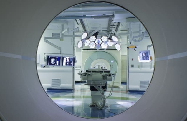
The Advanced Multimodality Image Guided Operating (AMIGO) suite, where procedures are performed, is a clinical translational test-bed for research of the National Center for Image-Guided Therapy.
The cryoablation procedure often begins by using CT guidance to drill into the bone for cryoprobe placement. The authors note that MRI can be used alone to place the cryoprobe if bone along the necessary path has already been completely eroded. Once the cryoprobe is in position, the team employ MRI to monitor growth of the cryoablation zone using standard axial and sagittal T2-weighted MR images. These can be acquired as rapidly as every 0.5 s, although are usually tailored to 1 min sequences in a 3T MRI.
Treatment comprises an initial cryoablation freeze for approximately 10 min, followed by a 5-min active thaw and a second freeze. The total procedure time varies between a couple of hours to an entire day depending on the complexity of the cancer. “Much of the time is in preparing the patient for the operating room and placing the cryoablation probe to avoid critical structures,” Lee explains. “This is usually an outpatient procedure, and patients can leave the hospital several hours after its completion.”

Lee and his team have now performed 11 MRI-guided cryoablation procedures for spinal canal decompression, and a total of 76 MRI-guided cryoablations in the head, neck and spine. He believes that Brigham and Women’s Hospital is the only hospital in the world currently performing MRI-guided cryoablation for spinal canal decompression.
“In addition to disease affecting the spinal cord, we have started treating disease affecting the brain,” he says. “It is an exciting new field where disease affecting the brain or spinal cord and minimally invasive treatment can be seen together on MRI to help patients. Basically, people about to be paralysed by cancer can now have hope that their cancer can be frozen with a needle, and that they can walk out the same day knowing that within the next month the cancer will die and their spine will regenerate on its own.”


