Using MRI and PET independently, researchers have further established that there is a significant relationship between excessive iron accumulation and tau protein deposits in brain regions linked to Alzheimer’s disease, according to a study published in Brain.
Quantitative susceptibility mapping (QSM) on MRI and tau-PET imaging showed significantly higher levels of iron and tau deposition, respectively, in subjects who were cognitively impaired and who tested positive for beta-amyloid protein, which also is associated with the onset of dementia.
“Together, these results provide in vivo evidence of an association between iron deposition and both tau aggregation and neurodegeneration, which help advance our understanding of the role of iron dysregulation in the Alzheimer’s disease aetiology,” wrote the authors, led by Nicola Spotorno, from Lund University and the University of Pennsylvania Perelman School of Medicine.

Recent neuroimaging studies have linked iron accumulation, as measured by QSM-MRI, with beta-amyloid accumulation and cognitive decline, as well as other neurological conditions, such as Parkinson’s disease. However, in vivo studies exploring a possible relationship between iron accumulation and tau deposits are scant.
“Considering neuronal iron accumulation has been associated with both normal aging and neurodegenerative processes, it follows that understanding the interaction between abnormal iron homeostasis and abnormal tau aggregation in Alzheimer’s disease could provide new insights into Alzheimer’s disease pathogenesis and lead to new therapeutic targets,” the authors wrote.
To delve deeper into this realm, Spotorno and colleagues took 236 participants, all of whom were beta-amyloid positive, from the Swedish BioFINDER-2 study. Of those subjects, 78 people (mean age, 78 years) were cognitively normal and 158 individuals (mean age, 78 years) were cognitively impaired, 81 of whom (51%) were diagnosed with Alzheimer’s disease.
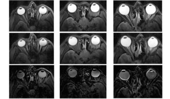
Participants underwent 3-tesla QSM-MR imaging (Prisma, Siemens Healthineers) to assess the extent of iron accumulation in grey matter, while PET imaging (Discovery 690, GE Healthcare) was performed with the tau tracer 18F-RO-948, which has succeeded in detecting tau protein tangles with little or no uptake in cognitively normal controls.
Comparisons of iron accumulation on QSM-MRI and tracer uptake on tau-PET in various brain regions confirmed a statistically significant difference between cognitively normal subjects and those with cognitive impairment, especially in the inferior temporal gyrus, which is “known to be severely affected by tau pathology in Alzheimer’s disease,” the authors wrote.

Interestingly, the difference between the two groups in QSM and tau-PET results was even more pronounced in beta-amyloid-positive individuals ages 65 and younger. In addition, both a voxel-wise analysis and region-of-interest-based analysis confirmed the associations between QSM and tau-PET with the “stereotypical spread of Alzheimer’s disease,” the authors added.
“These results advance our understanding of the role of iron dysregulation in Alzheimer’s disease pathophysiology and prompt new avenues for both research and treatment development work,” Spotorno and colleagues concluded.
- This article was originally published on AuntMinnie.com. ©2020 by AuntMinnie.com. Any copying, republication or redistribution of AuntMinnie.com content is expressly prohibited without the prior written consent of AuntMinnie.com.