A new nanothermometer that could be used to measure temperature variations in living cells has been created by researchers at Harvard University in the US. The device, which is based on diamond nanocrystals and is “injected” into the interior of cells using nanowires, can detect temperature fluctuations as small as 1.8 millikelvin over nanometre length scales. If further improved, it could be used to probe a range of temperature-sensitive phenomena in biological cells, and might even help in the development of “thermoblative” cancer treatments.
In their new work, the researchers, led by Mikhail Lukin of Harvard University, exploited nitrogen vacancy (NV) centres in diamond. NVs are defects that occur when two neighbouring carbon atoms are replaced by a nitrogen atom and an empty lattice site.
The ground state of an NV centre is split into two energy levels – low and high – and the energy difference between the two levels is known as the transition frequency. When diamond cools or warms, the NV transition frequency shifts accordingly. The new nanothermometry technique works by accurately measuring this shift – which can be detected using fluorescence spectroscopy – and then using this measurement to calculate the exact temperature of the nanodiamond. And since diamond is a good conductor of heat, it is likely to have the same temperature as its immediate surroundings – in this case a biological cell.
“Thanks to the small size of an individual NV centre, diamonds just a few tens of nanometres in size can be employed in the measurements,” explains team member Peter Maurer, who is a member of Lukin’s group. “This allows for a highly sensitive thermometer that is also compatible with the size of living cells, which themselves are microns in diameter.”
The device, which the researchers injected into biological cells during their experiments using nanowire “needles”, can detect temperature variations as small as 1.8 mK (in an ultra-pure bulk-diamond sample) and over distances as short as 200 nm.
Thermoblative therapy for tumours?
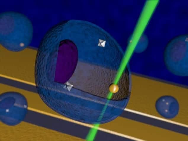
In another set of experiments, Lukin and colleagues combined their nanodiamond thermometer with gold nanoparticles that had been excited with laser light and so acted as localized heat sources. This technique allowed the team to both monitor and control the temperature in a biological cell – in one particular case a single human embryonic fibroblast. The heat generated by the gold nanoparticles could also be used to destroy the cell, the researchers found. Indeed, they succeeded in calculating the exact amount of heat required to do this. “We believe that combining such ‘thermoblative’ therapy with our temperature nanosensor could be a powerful tool for selectively identifying and killing malignant tumour cells, for example, without damaging surrounding healthy tissue,” says team member Georg Kucsko.
“If we further improve the sensitivity of our device, we could use it to observe real-time nanoscale processes in biological cells,” he told physicsworld.com. “We might also use it to directly monitor and control biochemical reactions in these cells. And combining our nanothermometer with techniques such as ‘two-photon’ microscopy may even allow us to identify local tumour activity in vivo by being able to map thermogenesis at the single-cell level.” Thermogenesis – the way heat is produced in living organisms – is different in tumour cells compared with healthy ones and this difference is one of the hallmarks of a tumour.
The research is published in the journal Nature.


