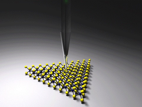
The promise of optical imaging and its wealth of spectroscopic information at nanoscale resolution seems too good to be true, and for many labs it has been. Diffraction limits the resolution of conventional optical microscopy to around half the wavelength of the illuminating light – roughly 100s of nanometres for visible light. Although scanning near-field optical microscopy (SNOM) beats the diffraction limit, as Ming Liu and Ruoxue Yan and colleagues at the University of California at Riverside (UCR) point out in a recent report, the spiralling sophistication of tip, instrumentation and optical designs to get the technique to work well can take its toll on the technique’s versatility and accessibility. Their report suggests a lens-free set up to optimize the efficiency of the technique, which could make it accessible even for labs limited to basic scanning probe apparatus.
Other techniques, such as scanning tunnelling and atomic force microscopy, have been capable of a resolution far beyond the diffraction limit since the 1980s. However, imaging with light gives features spectroscopic detail from interactions like Raman scattering, an extra dimension to the resulting images akin to moving from black and white to technicolour. The interactions between light and vibrational modes in molecules that leave their mark on light scattered from a sample – Raman scattering – reveal such a level of detail about structures and their environments they are often described as the sample’s “fingerprint”. By incorporating their SNOM set up – essentially a silver nanowire and an optical fibre coated in gold, both tapered at the tips – Liu, Yan and colleagues have brought this technicolour imaging capability to a standard teaching-level scanning tunnelling microscope.
Coupling issues
SNOM gets around the diffraction limit by measuring the “near-field”, the component of light that hugs surfaces, instead of the diffraction-prone “far-field” light that propagates away from structures that scatter it. To capture this non-propagating near-field, one approach has been the use of optical fibres brought within nanometres of the surface. However, for fibres with sufficiently narrow ends to extract nanoscale resolution information, getting light down the fibre to the sample and back in again presents its own challenges.
“Sending light through a tiny pinhole a thousand-times smaller than the diameter of a strand of human hair is no piece of cake,” Liu said. “Only a few in a million photons, or light particles, can pass the pinhole and reach the object you want to see. Getting a one-way ticket is already challenging; a round-trip ticket to bring back a meaningful signal is almost a daydream.”
The challenge has prompted interest in “apertureless” SNOM, where a metal nanoscale tip scatters the near-field at the sample surface to collect the high-resolution optical imaging data. However the level of background noise to signal can still make it difficult to obtain high-quality nanoscale information with apertureless SNOM, resulting in elaborate set ups and procedures to make the technique work well, despite the use of materials and geometries to exploit near-field enhancements through lightning rod and “plasmon resonance” effects.
Plasmons describe the way electrons in some metals respond to incident electromagnetic fields in unison at resonant wavelengths. SNOM researchers often exploit photons coupled to these oscillating resonant electron excitations at metal surfaces – “surface plasmon polaritons” – to achieve highly localized concentrated electromagnetic fields that enhance interactions between samples and light to get better optical measurements. However large differences in wavenumber can make it difficult to couple the far-field propagating light and the highly confined localized-surface-plasmon mode.
Thinning out the differences
To tackle the wave number mismatch, the UCR researchers exploit the gradual decrease in the effective mode index of the fibre as it tapers, and the resulting increase in wavelength, which is inversely proportional to this index. “The wavelength of the far-field light slowly increases as it travels down a gradually thinning optical fibre, without changing its frequency,” says Yan. “When it matches the wavelength of the electron density wave in the silver nanowire lying on top of the optical fibre, boom! All energy is transferred to the electron density wave and starts to travel on the surface of the nanowire instead.”
The researchers identify what they describe as “the only mode without cutoff and that can be effectively focused on the apex of a tapered rod”, which is, the radial transverse magnetic fundamental mode TM0. They then use linearly polarized light in the optical fibre, which will couple to that mode. The nanowire then tapers to just a few nanometres at the end to allow measurements with nanometre resolution. As well as increasing the intensity at the nanowire apex, effectively coupling this optical fibre light to TM0 SPP minimizes the background illumination.
Plasmonics technologies take on global challenges
Yan tells Physics World how as a graduate student in Peidong Yang’s group she worked on a device called a “nanowire endoscope”, which uses a tapered optical fibre to couple light into a tin oxide nanowire waveguide to guide visible light into intracellular compartments of a living mammalian cell. “The idea of coupling light to a plasmonic waveguide for tighter mode confinement and E-field enhancement for spectroscopy was sparked by the endoscopy work,” says Yan, emphasizing the challenge that bridging the momentum gap posed. “It was a long learning curve to understand this coupling system to single out the coupling condition for the high-efficiency excitation for the TM0 mode to synthetically modify to the tip morphology of the nanowire for high-resolution imaging, and to piece them all together.”
The researchers incorporate the approach on a standard scanning tunnelling microscope used for teaching, and image carbon nanotubes with 50% efficiency – 70% each for the fibre -nanowire-fibre coupling and for funnelling the light at the end of the nanowire.
They conclude in their report, “By offering an easy solution for efficiency light injection and/or extraction at a nanometre length scale, fibre-based near-field nanoscopy holds great potential as a plugin module for existing high-resolution measurement platforms to provide complementary and spatially correlated information on molecular compositions (for example, TERS), material properties (for example, inter- and intraband transitions) and optoelectronic device performance (for example, photocurrent mapping).”
Full details of Liu and Yan’s work alongside colleagues including Sanggon Kim, Ning Yu, Xuezhi Ma, Yangzhi Zhu, and Qiushi Liu are available in Nature Photonics.


