Stimulation of angiogenesis — the growth of new blood vessels — can be used to treat heart disease or promote wound healing. Meanwhile, inhibition of angiogenesis can be used as a therapeutic for cancer, ophthalmic conditions and other diseases. The delivery of proangiogenic therapeutics is thus a significant area of interest in the drug delivery research field. In this context, synthetic 2D nanomaterials are emerging as ideal structures for regenerative medicine applications, due to their biocompatibility and homogeneous physical and chemical characteristics.

With this in mind, a team of researchers from Texas A&M University are investigating the use of 2D nanosilicates as a platform technology to deliver proangiogenic growth factors to stimulate angiogenesis. This platform has the potential to be broadly used for growth factor delivery and release (Adv. Biosys. 10.1002/adbi.201800092).
Nanosilicates are 2D disc-shaped nanoparticles that interact with biomolecules. The interaction is electrostatic and results in the adsorption of the molecules on the surface of the nanosilicates. The authors confirmed this by adding proteins into a nanosilicate solution. The results suggested that the proteins adsorbed onto the nanosilicates and were released slowly over a course of weeks.
Next, the authors used a 3D invasion assay to examine the effect of growth factor-loaded nanosilicates on the sprouting step of angiogenesis. They did this by including proangiogenic proteins (such as VEGF, FGF and PDGF) to stimulate the invasion of endothelial cells into a basal 3D collagen matrix (the nanosilicates were incubated with growth factors and then mixed into collagen matrices).
The results indicated that the minimum concentration of nanosilicates required to sequester growth factors is 0.015%, and that at this concentration, the cells penetrated the collagen matrices and formed sprouting structures.
The researchers also tested the effect of nanosilicates on the mechanical properties of collagen matrices, concluding that a low concentration of nanosilicates (0.015%) does not affect the mechanical properties of the matrices. Thus, they examined the effects of nanosilicates in the 3D collagen invasion system, observing that growth factors appear to release from the nanosilicates quickly and were homogenously localized throughout the collagen matrix. These results indicate the ability of the nanosilicates to deliver angiogenic factors in specific combinations and efficiencies to direct cellular invasion.

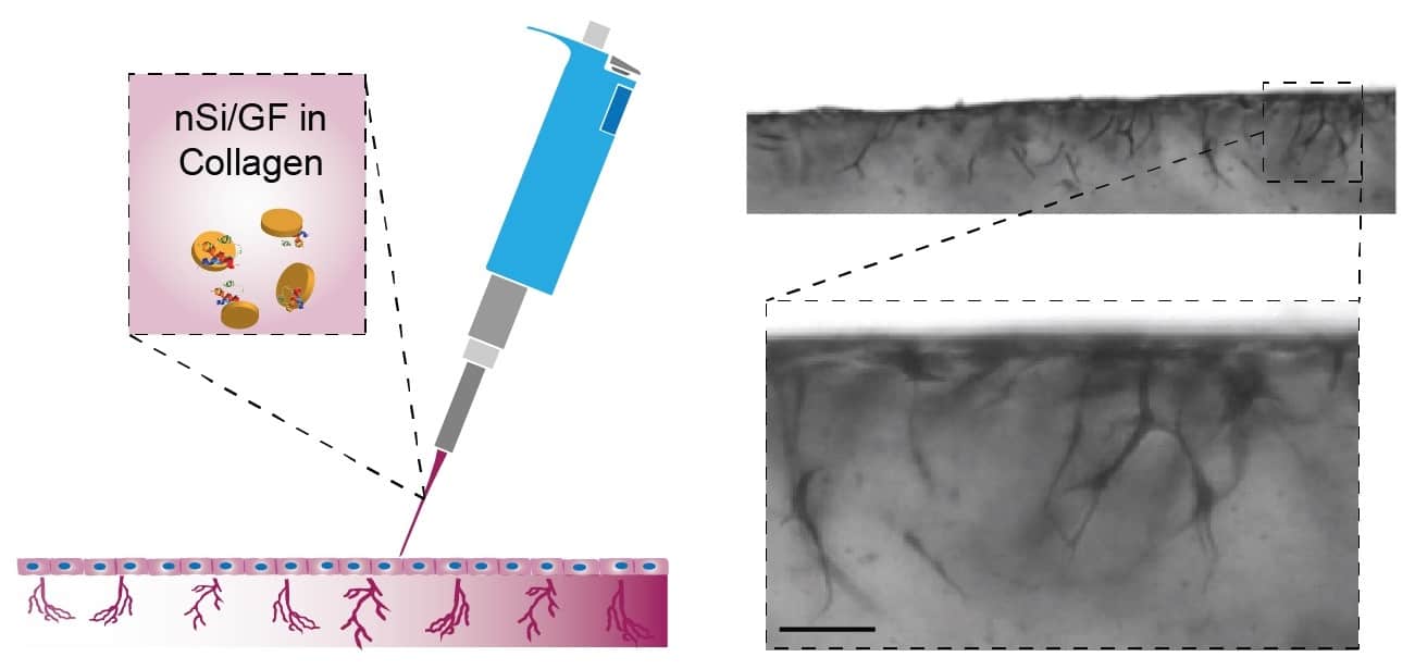
In another interesting development, the authors fabricated injectable collagen-based scaffolds that could be patterned by the inclusion of growth factors, and observed the invasion of endothelial cells. This method has potential applications in tendon or ligament repair.
The study demonstrates the ability of nanosilicates to deliver growth factors to induce an angiogenic response. The results also show the huge potential of nanosilicates for delivering biomolecules, thus paving the way for new therapeutics.


