Physicists have long been pioneers in medical science, from the earliest diagnoses with X-rays to the more recent development of techniques such as positron emission tomography, magnetic resonance imaging (MRI) and hadron therapy. In a series of three new videos, our colleagues at medicalphysicsweb discuss some recent exciting developments in medical physics with leading lights in the field.
First is an interview with Simon Cherry from the University of California, Davis, who is the incoming editor-in-chief of the journal Physics in Medicine and Biology. Founded in 1956, the journal’s previous editors include Nobel laureate Joseph Rotblat, who famously quit the Manhattan atomic-bomb project on humanitarian grounds and went on to have a starring career in medical physics.
In the interview, Cherry focuses on the benefits of “molecular imaging”, which can pinpoint the biochemical and molecular changes that accompany the very early stages of chronic diseases such as cancer or neurodegenerative diseases such as Alzheimer’s. As Cherry explains, such information is impossible to obtain with traditional clinical-imaging techniques such as X-ray or MRI, which largely reveal structural changes in the human body.
Cherry also explains how the Cerenkov effect – a well-established physical phenomenon – is now being exploited within the medical arena. The effect occurs when certain radionuclides, in addition to emitting gamma rays, also give off charged particles that, temporarily at least, travel through tissue faster than light in that medium. The particles emit characteristic “Cerenkov radiation” that can be used for imaging purposes. “Cerenkov luminescence imaging” is particularly useful for radionuclides such as yttrium-90 that do not emit any gamma rays and so are not easy to image by other means.
In the second interview, Uwe Oelfke from the German Cancer Research Center in Heidelberg, explains how image-guided radiation therapy (IGRT) can address one of the key challenges in modern radiotherapy – namely how to deliver a lethal dose of radiation to a tumour while sparing surrounding healthy tissue. The problem is that radiotherapy generally involves directing an invisible beam at an invisible tumour, based on patient images acquired prior to the treatment. Oelfke explains how IGRT involves acquiring additional images of the patient in the treatment position, immediately before or during radiation treatment, ensuring that the beam is precisely targeted at the tumour.
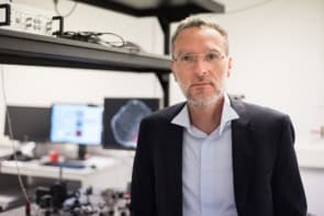
Finally, Freek Beekman, head of radiation, detection and medical imaging at the Delft University of Technology in the Netherlands, discusses the use of a technique known as “single-photon emission computed tomography” (SPECT), which can create 3D images of the distribution of gamma rays emitted by a radionuclide. SPECT is particularly useful for imaging small animals, which is invaluable for developing new therapeutic strategies for human medicine, offering, for example, a way of tracking the response of pharmaceuticals in animal models of cancer, diabetes and other diseases. Beekman also discusses the work of MILabs, a company that he founded to commercialize pre-clinical molecular imaging and that offers a range of SPECT systems.