Electron microscopy is unique in its ability to provide high-resolution cellular imaging, but it does not give information about the location of specific proteins in a cell. Labelling with electron-dense particles like gold enables visualization of proteins, but is limited to one type of protein at a time. To overcome this limitation, a group of researchers in the US have presented nine nanoparticles with distinct colours that might allow multicolour electron microscopy in the future (Nature Nanotechnol. 10.1038/s41565-019-0395-0).
Maxim Prigozhin, Peter Maurer and colleagues made nanoparticles from NaGdF4 or NaYF4 and spiked them with one of nine lanthanide ions: Eu3+, Er3+, Ho3+, Tb3+, Sm3+, Dy3+, Nd3+, Tm3+ and Yb3+. These rare-earth elements determine the colour of the nanoparticle via a phenomenon called cathodoluminescence. The resulting nanoparticles represent the first ever coloured labels for electron microscopy.

What is cathodoluminescence?
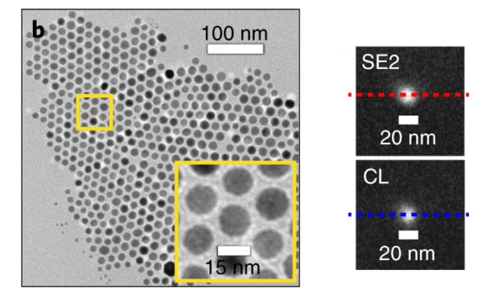
Cathodoluminescence is a phenomenon in which electrons impacting on a material, in this case the lanthanide-spiked nanoparticles, emit light of material-specific wavelengths. In this study, the electron beam of the electron microscope was used to excite the nanoparticles. The resulting light emission spectra of the nine types of nanoparticle were distinct, making them a potential stepping stone towards multicolour imaging.
The researchers also tested three additional lanthanides, which did not yield sharp spectra like the other nine elements. A number of lanthanides remain to be tested and might yield further colours to expand the repertoire. To create even more colours, the researchers are also thinking about co-doping nanoparticles with multiple lanthanides.
The resulting nanoparticles had diameters of less than 20 nm, which is comparable to the quantum dots, gold nanoparticles and immunoglobulin antibodies typically used to label proteins in electron microscopy. The big advantage of the new nanoparticles is that they come in nine different colours, such that co-labelling of several proteins might be possible. At the same time, cathodoluminescence-electron microscopy images of the new nanoparticles showed that this method is suitable for nanoscale imaging.
The main problem that the researchers encountered was variability between experiments in the level of the coloured signal emitted from the nanoparticles. However, they are confident that this will be manageable with further optimization.
Future potential
The team, led by Nobel Laureate Steven Chu, outline many ways in which their method can be optimized. These include further reducing the nanoparticle size, optimizing the nanoparticle surface and making use of the long-excited state of the lanthanides to distinguish signal from noise by time-gating the measurements. Combined, these approaches might allow the design of even smaller labels for biological multicolour electron microscopy. Another way to improve the signal-to-noise ratio might be to reduce the energy of the electron beam, thereby matching it to the size needed to excite the nanoparticle.
In the future, the researchers envision that instead of scanning the whole sample for cathodoluminescence, imaging might be accelerated by identifying the nanoparticles through conventional electron microscopy and then only using cathodoluminescence to identify the colour of each nanoparticle.