Biophysics

Biophysics

Non-destructive electron microscopy maps amino acids

28 Feb 2019 Lucy Rowlands 
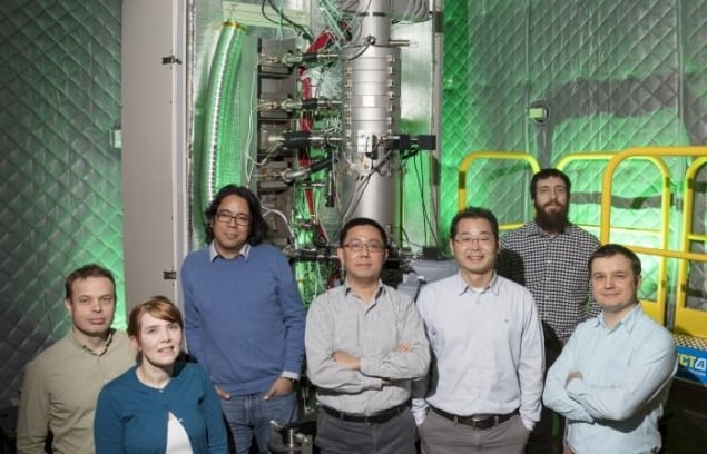
Oak Ridge team
The team from Oak Ridge National Laboratory, with the electron microscope that can identify site-specific isotopic labels. (Courtesy: Carlos Jones/Oak Ridge National Laboratory, US Dept of Energy)

Researchers from Oak Ridge National Laboratory have developed an electron microscopy technique that can detect different isotopes in amino acids. The non-destructive technique means that scientists can spatially track amino acids, enabling access to unprecedented details of biological processes. This is particularly useful in the study of protein interactions, which can shed light on disease progression and other complex biological events (Science 10.1126/science.aav5845).

Traditionally, to study protein interactions, scientists label the protein-of-interest with a specific isotope and watch how its mass changes. Current methods, such as mass spectrometry and optical techniques, are mainly useful at the macroscopic level and can destroy the sample in the process. This new type of microscopy will allow scientists to follow the interaction through space and map the location of labelled amino acids while leaving them intact.

New vibrations

The research team used monochromated electron energy-loss spectroscopy (EELS) in conjunction with a scanning transmission electron microscope (STEM). With this technique, a negatively charged electron beam is positioned very close to the sample so that the beam only grazes over it. This means that the machine can both excite and detect molecular vibrations without destroying the sample.

Usually, the negatively charged electron beam used for electron microscopy is only sensitive to protons. However, the frequency of the molecular vibration is also dependent on atomic mass, which means that heavier isotopes shift the vibrational modes. These shifts were then used by the team to track the labelled amino acids.

The researchers were able to distinguish between amino acids labelled with carbon-12 and carbon-13 with nanoscale spatial resolution. They then used this method to look at crystals of alanine and map the distribution of the labelled acids throughout the structures.

Working with mass spectrometry

Protein labelling is usually performed using mass spectrometry, which has excellent sensitivity, but destroys the sample in the process, thus losing key information about atom connections. Therefore, the information extracted is only a snapshot of one moment in time. The authors of this study don’t believe that their new technique will replace mass spectrometry, but instead suggest that it will offer a complementary method.

“Our technique is the perfect complement to a macroscale mass spectrometry experiment,” says lead author Jordan Hachtel. “With the pre-knowledge of the mass spectrometry, we can go in and spatially resolve where the isotopic labels are ending up in a real-space sample.”

The technique could also have applications in areas such as research into polymers and other soft matter. It could find particular use in the field of quantum materials, where isotopic substitution is critical to control superconductivity.

Copyright © 2024 by IOP Publishing Ltd and individual contributors