Measuring blood flow to the brain is essential for diagnosing and developing treatments for neurological disorders such as stroke, vascular dementia or traumatic brain injury. Performing this measurement non-invasively is challenging, however, and achieved predominantly using costly MRI and nuclear medicine imaging techniques.
Emerging as an alternative, modalities based on optical transcranial measurement are cost-effective and easy to use. In particular, speckle contrast optical spectroscopy (SCOS) – an offshoot of laser speckle contrast imaging, which uses laser light speckles to visualize blood vessels – can measure cerebral blood flow (CBF) with high temporal resolution, typically above 30 Hz, and cerebral blood volume (CBV) through optical signal attenuation.
Researchers at the California Institute of Technology (Caltech) and the Keck School of Medicine’s USC Neurorestoration Center have designed a lightweight SCOS system that accurately measures blood flow to the brain, distinguishing it from blood flow to the scalp. Co-senior author Charles Liu of the Keck School of Medicine and team describe the system and their initial experimentation with it in APL Bioengineering.
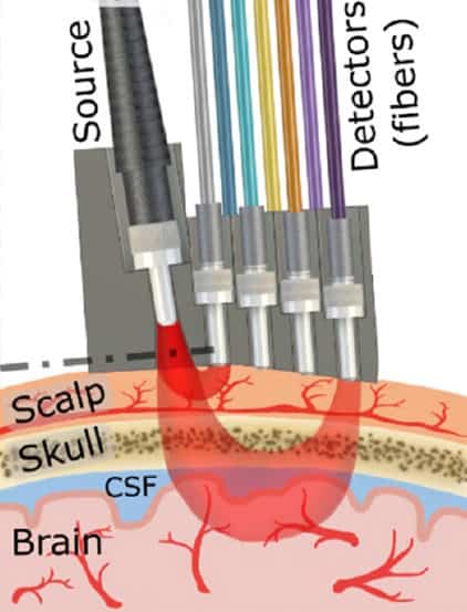
The SCOS system consists of a 3D-printed head mount designed for secure placement over the temple region. It holds a single 830 nm laser illumination fibre and seven detector fibres positioned at seven different source-to-detector (S–D) distances (between 0.6 and 2.6 cm) to simultaneously capture blood flow dynamics across layers of the scalp, skull and brain. Fibres with shorter S–D distances acquire shallower optical data from the scalp, while those with greater distances obtain deeper and broader data. The seven channels are synchronized to exhibit identical oscillation frequencies corresponding to the heart rate and cardiac cycle.
When the SCOS system directs the laser light onto a sample, multiple random scattering events occur before the light exits the sample, creating speckles. These speckles, which materialize on rapid timescales, are the result of interference of light travelling along different trajectories. Movement within the sample (of red blood cells, for instance) causes dynamic changes in the speckle field. These changes are captured by a multi-million-pixel camera with a frame rate above 30 frames/s and quantified by calculating the speckle contrast value for each image.
Human testing
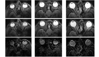
The researchers used the SCOS system to perform CBF and CBV measurements in 20 healthy volunteers. To isolate and obtain surface blood dynamics from brain signals, the researchers gently pressed on the superficial temporal artery (a terminal branch of the external carotid artery that supplies blood to the face and scalp) to block blood flow to the scalp.
In tests on the volunteers, when temporal artery blood flow was occluded for 8 s, scalp-sensitive channels exhibited significant decreases in blood flow while brain-sensitive channels showed minimal change, enabling signals from the internal carotid artery that supplies blood to the brain to be clearly distinguished. Additionally, the team found that positioning the detector 2.3 cm or more away from the source allowed for optimal brain blood flow measurement while minimizing interference from the scalp.
“Combined with the simultaneous measurements at seven S–D separations, this approach enables the first quantitative experimental assessment of how scalp and brain signal contributions vary with depth in SCOS-based CBF measurements and, more broadly, in optical measurements,” they write. “This work also provides crucial insights into the optimal device S–D distance configuration for preferentially probing brain signal over scalp signal, with a practical and subject-friendly alternative for evaluating depth sensitivity, and complements more advanced, hardware-intensive strategies such as time-domain gating.”
The researchers are now working to improve the signal-to-noise ratio of the system. They plan to introduce a compact, portable laser and develop a custom-designed extended camera that spans over 3 cm in one dimension, enabling simultaneous and continuous measurement of blood dynamics across S–D distances from 0.5 to 3.5 cm. These design advancements will enhance spatial resolution and enable deeper brain measurements.
Laser-based headset assesses stroke risk using the brain’s blood flow

“This crucial step will help transition the system into a compact, wearable form suitable for clinical use,” comments Liu. “Importantly, the measurements described in this publication were achieved in human subjects in a very similar manner to how the final device will be used, greatly reducing barriers to clinical application.”
“I believe this study will advance the engineering of SCOS systems and bring us closer to a wearable, clinically practical device for monitoring brain blood flow,” adds co-author Simon Mahler, now at Stevens Institute of Technology. “I am particularly excited about the next stage of this project: developing a wearable SCOS system that can simultaneously measure both scalp and brain blood flow, which will unlock many fascinating new experiments.”