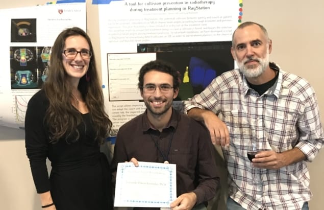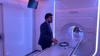
A team at Massachusetts General Hospital (MGH) has developed RadCollision – an open-source collision detection tool designed to aid dosimetrists planning photon or proton beam radiotherapy. When embedded in a treatment planning system (TPS), the modular software platform takes just seconds to automatically calculate whether a gantry head will collide with the patient or treatment couch. In addition to greatly reducing the need for replanning if a collision is detected during the dry run, the tool helps planners choose optimum and feasible irradiation angles to potentially increase the quality of treatment plans.
The MGH research initiative began when the radiotherapy department’s medical dosimetrists requested software to eliminate guesswork regarding collision risks. The dosimetrists previously relied on their experience and intuition to determine which incident beam directions and isocentre positions may be infeasible, a time-consuming and less than optimum process.
“The frequency of potential collisions in the clinic has been mitigated by rudimentary physical measurements and conservative planning decisions,” explains co-author Kyla Remillard, a photon dosimetry team leader. “Without these steps, potential collisions would be observed more frequently. However, no two patients’ geometries or setups are identical, and so we say there’s an opportunity for improvement.”
Writing in Biomedical Physics & Engineering Express, Remillard and colleagues explain that equipment movement during photon treatments can be considerable, with the treatment head mounted on a rotating gantry and a patient couch rotating around a vertical axis. Movement during proton therapy is even greater, and therefore more complex to calculate, because the moving snout of the treatment head nozzle supports apertures, compensators and range shifters positioned close to the surface of a patient. Additionally, couches with robotic arms can lead to some extra risk of collision when the beam is incident from below.
Collision avoidance is based on a detailed 3D description of static objects in each treatment room, as well as the patient’s 3D anatomy, the authors explain. Current methods of assessing potential collisions include the use of 3D scanners or cameras, patient geometry data from a CT scan, and/or analytic software. These typically require setup-related costs, especially if a cancer treatment centre uses multiple types of treatment systems and patient couches. Some vendors include 3D visualization tools for real-time interaction with the delivery machine. But these are not usually embedded in the planning software and may also add licensing costs.
The free RadCollision software does not require purchase of additional software or hardware, and is easily adaptable by any institution for any type of treatment equipment configuration. RadCollision relies on an initial 3D modelling of the treatment machine, and optionally incorporates the full patient geometry recorded with any 3D scanner or surface imaging device. It provides a realistic 3D visualization of nozzle, couch and patient, and its modularity enables the user to add or remove room elements from the 3D visualization.

Other features include the ability to evaluate the independent movement of each treatment room element with real-time feedback, and interactive sliders that help planners choose optimal beam angles. Users also can choose automatic or visual detection modes, with the latter relying on the planner’s ability to assess the collision risk in incomplete patient geometries.
RadCollision can be used as soon as the patient’s contour data are added. The program automatically selects the machine and couch model from the active treatment plan. Dosimetrists may adjust the irradiation settings, after which, the software transforms in real time the regions-of-interest (ROIs) corresponding to the treatment machine, calculating any collision (overlap of ROIs) with the patient or couch. Collision reports can be automatically calculated for each beam defined in the plan.
The tool requires an initial setup performed by a hospital’s information technology department. The RadCollision software needs to be embedded into each TPS database (if more than one TPS is used) and a folder (STL files) with the 3D models of the machines prepared. RadCollision is currently limited to use with the RayStation TPS, but versions for use with other commercial TPS are planned, says first author Fernando Hueso-González.
The researchers quantitatively evaluated their software using the RayStation TPS with four patient treatment plans that were found infeasible during previous collision checks by therapists. The software reported collisions with the couch at similar angles to those reported experimentally. The team also tested the software with a model of a proton treatment room and a robotic patient positioning system.
“In one case, we tested in the RadCollision software a beam where the dosimetrist doubted that there was enough clearance with the toe of a patient’s foot. RadCollision predicted that clearance would be very tight, but the irradiation-optimized TPS was feasible,” comments Remillard. “When we performed a dry run, there was no collision.”
The team note that the reliability of the collision assessment depends upon the accuracy of the input data. Sources of uncertainty include changes in patient anatomy or position, patient motion, CT and/or 3D scan data, and the accuracy of 3D models of the machine and couch.
RadCollision has been created with the hope that it will aid in the development of optimally individualized treatment plans. “By providing a real-time assurance that the selected angles do not present a risk of collision, dosimetrists are less likely to shy away from irradiation geometries beneficial from the dose perspective,” write the authors. “RadCollision could be most helpful for clinical cases such as stereotactic treatments, extremities, partial breast irradiation and prone breast treatments, electron beams, and plans with drastically anterior or posterior isocentres.”
“We are aware of hospitals in the UK, France, Italy, the Netherlands, Spain and the United States trying out our script,” Hueso-González tells Physics World. “We can’t use it yet on a clinical basis here at Mass General because we are awaiting an upgrade of TPS software that will be compatible. We anticipate starting to use it clinically in January 2021.”


