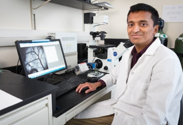
Narasimhan Rajaram, a biomedical engineer at the University of Arkansas, has received a $2.03 million grant to develop optical imaging technologies that identify therapy-resistant tumours early in the treatment process. The five-year grant, from the National Cancer Institute, will support the creation of a device to monitor the response of head-and-neck cancer patients to radiotherapy and chemotherapy during treatment.
The current standard-of-care for head-and-neck tumours involves a seven-week regimen of radiation and chemotherapy, followed by MRI and X-ray CT eight weeks after treatment to determine whether the tumour has responded.
“The long treatment duration makes it imperative to find out right away if changes are required to the treatment regimen for non-responding tumours,” says Rajaram. “Exceptional responders could also benefit by allowing potential de-escalation of the radiation dose. Unfortunately, there are currently no methods that can identify treatment response in the clinic during therapy, which causes patients – both responsive and resistant – to lose critical time when alternative approaches could be considered.”
To remedy this situation, Rajaram’s team has partnered with researchers from Johns Hopkins University and the University of Arkansas for Medical Sciences (UAMS). Technology development and pre-clinical studies will be conducted at the University of Arkansas and Johns Hopkins, while the clinical trials will be conducted at UAMS.
The researchers aim to develop an endoscope-compatible fibre-optic probe that combines diffuse reflectance spectroscopy and Raman spectroscopy. Diffuse reflectance spectroscopy uses optical fibres to deliver low-power, non-ionizing visible light onto tissue and collect the diffusely reflected light. The Rajaram lab has developed models of light–tissue interaction to extract quantitative information, such as tissue oxygenation, from this reflected light.
Raman spectroscopy predicts radiation resistance
Raman spectroscopy uses inelastic scattering of near-infrared laser light to provide a highly specific fingerprint of molecules in tissue. Since every molecule has unique Raman features, mathematical models can be used to identify and quantify the contributions of individual molecules.
“These complementary tools can provide information about tumour oxygenation levels, which is critical for radiation therapy to work, as well as the contributions of key biomolecules in the tumour microenvironment that contribute to the development of radiation resistance,” Rajaram explains.
He adds that the technologies developed in this project could also be used to evaluate other treatments, such as new drugs being developed to treat different cancers.


