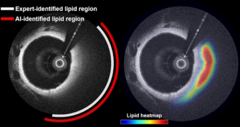
Optical coherence tomography (OCT), a low-cost imaging technology used to diagnose and plan treatment for eye diseases, also shows potential as a diagnostic tool for assessing rapid hearing loss.
Researchers at the Keck School of Medicine of USC have developed an OCT device that can acquire diagnostic quality images of the inner ear during surgery. These images enable accurate measurement of fluids in the inner ear compartments. The team’s proof-of-concept study, described in Science Translational Medicine, revealed that the fluid levels correlated with the severity of a patient’s hearing loss.
An imbalance between the two inner ear fluids, endolymph and perilymph, is associated with sudden, unexplainable hearing loss and acute vertigo, symptoms of ear conditions such as Ménière’s disease, cochlear hydrops and vestibular schwannomas. This altered fluid balance – known as endolymphatic hydrops (ELH) – occurs when the volume of endolymph increases in one compartment and the volume of perilymph decreases in the other.
Because the fluid chambers of the inner ear are so small, there has previously been no effective way to assess endolymph-to-perilymph fluid balance in a living patient. Now, the Keck OCT device enables imaging of inner ear structures in real time during mastoidectomy – a procedure performed during many ear and skull base surgeries, and which provides optical access to the lateral and posterior semicircular canals (SCCs) of the inner ear.
OCT offers a quicker, more accurate and less expensive way to see inner ear fluids, hair cells and other structures compared with the “gold standard” MRI scans. The researchers hope that ultimately, the device will evolve into an outpatient assessment tool for personalized treatments for hearing loss and vertigo. If it can be used outside a surgical suite, OCT technology could also support the development and testing of new treatments, such as gene therapies to regenerate lost hair cells in the inner ear.
Intraoperative OCT
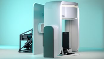
The intraoperative OCT system, developed by senior author John Oghalai and colleagues, comprises an OCT adaptor containing the entire interferometer, which attaches to the surgical microscope, plus a medical cart containing electronic devices including the laser, detector and computer.
The OCT system uses a swept-source laser with a central wavelength of 1307 nm and a bandwidth of 89.84 nm. The scanning beam spot size is 28.8 µm and has a depth-of-focus of 3.32 mm. The system’s axial resolution of 14.0 µm and lateral resolution of 28.8 µm provide an in-plane resolution of 403 µm2.
The laser output is directed into a 90:10 optical fibre fused coupler, with the 10% portion illuminating the interferometer’s reference arm. The other 90% illuminates the sample arm, passes through a fibre-optic circulator, and is combined with a red aiming beam that’s used to visually position the scanning beam on the region-of-interest.
After the OCT and aiming beams are guided onto the sample for scanning, and the interferometric signal needed for OCT imaging is generated, two output ports of the 50:50 fibre optic coupler direct the light signal into a balanced photodetector for conversion into an electronic signal. A low-pass dichroic mirror allows back-reflected visible light to pass through into an eyepiece and a camera. The surgeon can then use the eyepiece and real-time video to ensure correct positioning for the OCT imaging.
Feasibility study
The team performed a feasibility study on 19 patients undergoing surgery at USC to treat Ménière’s disease (an inner-ear disorder), vestibular schwannoma (a benign tumour) or middle-ear infection with normal hearing (the control group). All surgical procedures required a mastoidectomy.
Immediately after performing the mastoidectomy, the surgeon positioned the OCT microscope with the red aiming beam targeted at the SCCs of the inner ear. After acquiring a 3D volume image of the fluid compartments in the inner ear, which took about 2 min, the OCT microscope was removed from the surgical suite and the surgical procedure continued.
The OCT system could clearly distinguish the two fluid chambers within the SCCs. The researchers determined that higher endolymph levels correlated with patients having greater hearing loss. In addition to accurately measuring fluid levels, the system revealed that patients with vestibular schwannoma had higher endolymph-to-perilymph ratios than patients with Ménière’s disease, and that compared with the controls, both groups had increased endolymph and reduced perilymph, indicating ELH.
The success of this feasibility study may help improve current microsurgery techniques, by guiding complex temporal bone surgery that requires drilling close to the inner ear. OCT technology could help reduce surgical damage to delicate ear structures and better distinguish brain tumours from healthy tissue. The OCT system could also be used to monitor the endolymph-to-perilymph ratio in patients with Ménière’s disease undergoing endolymphatic shunting, to verify that the procedure adequately decompresses the endolymphatic space. Efforts to make a smaller, less expensive system for these types of surgical use are underway.
First human retinal image brings sight-saving portable OCT a step closer

The researchers are currently working to improve the software and image processing techniques in order to obtain images from patients without having to remove the mastoid bone, which would enable use of the OCT system for outpatient diagnosis.
The team also plans to adapt a handheld version of an OCT device currently used to image the tympanic membrane and middle ear to enable imaging of the human cochlea in the clinic. Imaging down the ear canal non-invasively offers many potential benefits when diagnosing and treating patients who do not require surgery. For example, patients determined to have ELH could be diagnosed and treated rapidly, a process that currently takes 30 days or more.
Oghalai and colleagues are optimistic about improvements being made in OCT technology, particularly in penetration depth and tissue contrast. “This will enhance the utility of this imaging modality for the ear, complementing its potential to be completely non-invasive and expanding its indication to a wider range of diseases,” they write.