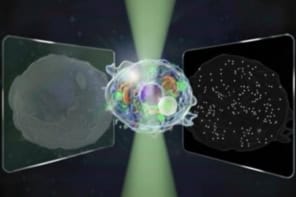
Physicists in Germany have used visible light to measure intramolecular distances smaller than 10 nm thanks to an advanced version of an optical fluorescence microscopy technique called MINFLUX. The technique, which has a precision of just 1 angstrom (0.1 nm), could be used to study biological processes such as interactions between proteins and other biomolecules inside cells.
In conventional microscopy, when two features of an object are separated by less than half the wavelength of the light used to image them, they will appear blurry and indistinguishable due to diffraction. Super-resolution microscopy techniques can, however, overcome this so-called Rayleigh limit by exciting individual fluorescent groups (fluorophores) on molecules while leaving neighbouring fluorophores alone, meaning they remain dark.
One such technique, known as nanoscopy with minimal photon fluxes, or MINFLUX, was invented by the physicist Stefan Hell. First reported in 2016 by Hell’s team at the Max Planck Institute (MPI) for Multidisciplinary Sciences in Göttingen, MINFLUX first “switches on” individual molecules, then determines their position by scanning a beam of light with a doughnut-shaped intensity profile across them.
The problem is that at distances of less than 5 to 10 nm, most fluorescent molecules start interacting with each other. This means they cannot emit fluorescence independently – a prerequisite for reliable distance measurements, explains Steffen Sahl, who works with Hell at the MPI.
Non-interacting fluorescent dye molecules
To overcome this problem, the team turned to a new type of fluorescent dye molecule developed in Hell’s research group. These molecules can be switched on in succession using UV light, but they do not interact with each other. This allows the researchers to mark the positions they want to measure with single fluorescent molecules and record their locations independently, to within as little as 0.1 nm, even when the dye molecules are close together.
“The localization process boils down to relating the unknown position of the fluorophore to the known position of the centre of the doughnut beam, where there is minimal or ideally zero excitation light intensity,” explains Hell. “The distance between the two can be inferred from the excitation (and hence the fluorescence) rate of the fluorophore.”
The advantage of MINFLUX, Hell tells Physics World, is that the closer the beam’s intensity minimum gets to the fluorescent molecule, the fewer fluorescence photons are needed to pinpoint the molecule’s location. This takes the burden of producing localizing photons – in effect, tiny lighthouses signalling “Here I am!” – away from the relatively weakly-emitting molecule and shifts it onto the laser beam, which has photons to spare. The overall effect is to reduce the required number of detected photons “typically by a factor of 100”, Hell says, adding that this translates into a 10-fold increase in localization precision compared to traditional camera-based techniques.
“A real alternative” to existing measurement methods
The researchers demonstrated their technique by precisely determining distances of 1–10 nanometres in polypeptides and proteins. To prove that they were indeed measuring distances smaller than the size of these molecules, they used molecules of a different substance, polyproline, as “rulers” of various lengths.
Polyproline is relatively stiff and was used for a similar purpose in early demonstrations of a method called Förster resonance energy transfer (FRET) that is now widely used in biophysics and molecular biology. However, FRET suffers from fundamental limitations on its accuracy, and Sahl thinks the “arguably surprising” 0.1 nm precision of MINFLUX makes it “a real alternative” for monitoring sub-10-nm distances.
While it had long been clear that MINFLUX should, in principle, be able to resolve distances at the < 5 nm scale and measure them to sub-nm precision, Hell notes that it had not been demonstrated at this scale until now. “Showing that the technique can do this is a milestone in its development and demonstration,” he says. “It is exciting to see that we can resolve fluorescence molecules that are so close together that they literally touch.” Being able to measure these distances with angstrom precision is, Hell adds, “astounding if your bear in mind that all this is done with freely propagating visible light focused by a conventional lens”.
Molecular measuring stick could advance super-resolution microscopy
“I find it particularly fascinating that we have now gone to the very size scale of biological molecules and can quantify distances even within them, gaining access to details of their conformation,” Sahl adds.
The researchers say that one of the key prerequisites for this work (and indeed all super-resolution microscopy developed to date) was the sequential ON/OFF switching of the fluorophores emitting fluorescence. Because any cross-talk between the two molecules would have been problematic, one of the main challenges was to identify fluorescence molecules with truly independent behaviour – that is, ones in which the silent (OFF-state) molecule did not affect its emitting (ON-state) neighbour and vice versa.
Looking forward, Hell says he and his colleagues are now looking to develop and establish MINFLUX as a standard tool for unravelling and quantifying the mechanics of proteins.
The research is published in Science.


