Radiation therapy plays a fundamental role in cancer treatment, but there is a global shortage of radiotherapy centres, with many low-to-middle-income countries having limited or no treatment capability. This situation exists in part due to the cost of facilities and the expense of acquiring and operating radiotherapy systems. Linear accelerators with simplified designs, such as fixed gantry systems, could reduce these costs.
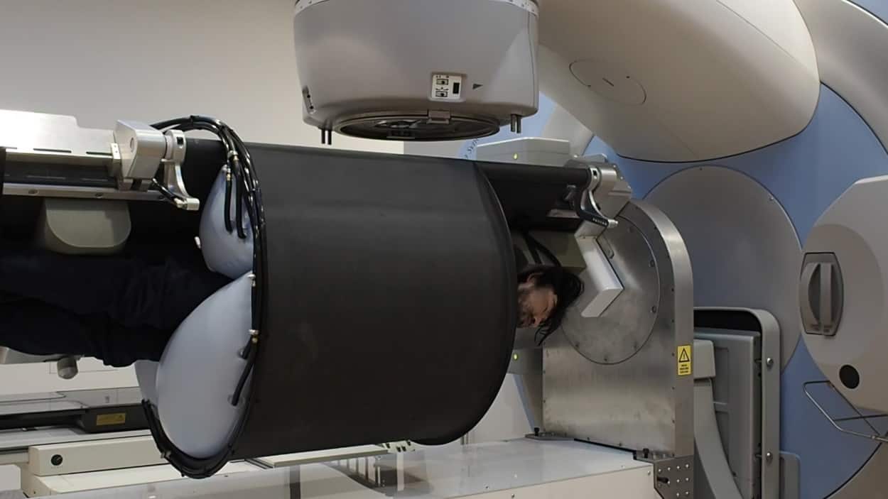
Researchers at the ACRF Image X Institute at the University of Sydney are developing a 3D conformal radiotherapy system with a fixed vertical X-ray beam, horizontal patient rotation and image guidance. The full-size proof-of-concept prototype, which offers high-quality radiation therapy from a smaller, more robust and more cost-effective system, has now been successfully commissioned (Med. Phys. 10.1002/mp.13356).
From a financial perspective, there are many potential advantages of a such a fixed-beam system. Without a rotating gantry, the system has fewer moving parts, which could improve reliability and robustness, and potentially reduce maintenance costs. It would also require less bunker shielding to operate safely, thereby reducing the cost of building new bunkers or renovating bunkers housing older radiotherapy equipment such as cobalt-60 units.
The prototype system — developed by Paul Liu and colleagues working on the Nano-X project to improve global access to radiotherapy — is based on the concept of patient rotation, specifically, keeping the radiation beam stationary while still achieving the necessary beam angles to achieve a desired dose distribution. Image guidance technologies will identify the tumour and adapt the treatment in real-time to ensure that the radiation dose is precisely delivered to the target.
The prototype comprises a standard Synergy linac with the gantry fixed at 0° and a horizontal patient rotation system (PRS). The PRS is a custom-designed radiotherapy couch equipped with straps for the head, chest, hips and legs, plus three independently controlled airbags that inflate over a patient’s chest and sides. The couch can move with two degrees-of-freedom to position and rotate the patient.

After the patient is immobilized and in a specified treatment position, they can be rotated to a specific angle for either kilovoltage (kV) imaging via the on-board imager or treatment with the megavoltage beam. The software operating the PRS allows for precise motion control, setting the target position or angle along with the desired velocity, acceleration and deceleration. It can also follow a series of queued motion commands, or execute quick-stop, return-to-home and patient egress commands.
The system passed all commissioning steps, which involved verification of geometric and dosimetric accuracy following conventional radiotherapy guidelines. The team also performed thorough testing of safety and interlock systems.
Clinical potential
The authors note that three essential steps will be needed before treating patients. Cone-beam CT image reconstruction under gravitational deformation may require advanced image reconstruction algorithms. They also need to develop methods to shift the beam to account for gravitational deformation-induced target motion.
Additionally, a patient’s tolerance of, and anxiety level relating to, horizontal rotation is unknown. It could be as much of a problem as an MRI exam is to a claustrophobic or noise-averse patient. An upcoming clinical trial will investigate and quantify how patients respond to strap and airbag immobilization and horizontal rotation.
Liu discussed the challenges with Physics World. “While we initially focused on static targets, an important part of the system will be its ability to adapt to motion, both from the patient’s normal physiological functions like breathing and from gravity as the patient is rotated,” he explains. “The next stage of the project will focus on implementing and testing algorithms that we’ve developed to both identify the amount of motion and to compensate for it accordingly.”
To enable real-time image guidance, the researchers are testing kilovoltage intrafraction monitoring (KIM), a novel tumour localization system developed at the University of Sydney that accurately estimates the 3D position of a target based on the 2D position of segmented markers in kV projections.
Real-time image-guided ART achieved on a standard linac
“KIM will offer real-time 3D target tracking with sub-degree and sub-millimetre accuracy,” Liu says. “We have successfully tested KIM together with real-time multileaf collimation tracking on a miniature version of this system, and are currently scaling these algorithms to our full-size prototype. We will be using KIM with a deformable phantom where the target will move as it undergoes rotation.”
The researchers are also investigating intensity-modulated radiotherapy and volumetric-modulated arc therapy, which are under various stages of implementation. Liu says that both are technically feasible, because the software and hardware control of the PRS has sufficient precision and flexibility.
Much work, followed by testing with veterinary radiation treatments, will be required before the first palliative treatments on human cancer patients can be undertaken. The system is not designed for infants, very small children or obese patients. But for all other cancer patients, this prototype radiotherapy system has potential to fill the existing and expanding gap between available treatment and need, especially for patients living in economically challenged areas of the world.


