Gamma imaging is a nuclear medicine technique employed in over 100 different diagnostic procedures. Also known as scintigraphy, the approach uses gamma cameras to image the distribution of gamma-emitting radiopharmaceuticals administered to the body, with applications including thyroid imaging, tumour imaging, and lung and renal studies.
Most clinical gamma cameras are large devices designed for whole-body scanning and located in their own dedicated room. While such systems offer high sensitivity and a large field-of-view (FOV), they are not ideal for patients who cannot attend the nuclear medicine department. A small gamma camera, on the other hand, could enable scanning of more patients in far more scenarios.
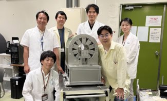
Made to meet this challenge, Seracam is a new portable gamma camera developed and designed by UK medical imaging company Serac Imaging Systems. Just 15 cm in diameter, 24 cm long and weighing 5 kg, the hybrid optical–gamma camera is designed for small-organ imaging within outpatient clinics, intensive care units, or even in operating theatres during surgery.
“Gamma imaging is no longer confined to the nuclear medicine department,” explains Sarah Bugby from Loughborough University. “The system has a much smaller footprint, it could be stored in a cupboard and brought out only when needed.”
Bugby and colleagues have now performed a detailed assessment of Seracam’s clinical potential, reporting their findings in EJNMMI Physics.
System evaluation
Seracam uses a CsI(Tl) crystal scintillator to convert incoming gamma photons to optical photons. This light is then captured by a 25.5 x 25.5 mm detector, divided into 245 x 245 pixels, and analysed in real time to create an image of the gamma counts.

The camera integrates four pinhole collimators, with pinhole diameters of roughly 1, 2, 3 and 5 mm. The physics of collimation means that smaller pinhole diameters provide better spatial resolution but lower sensitivity, while larger pinholes provide higher sensitivity but with a trade-off in image resolution.
“With Seracam, you can change the collimator at the press of a button in just a second or so,” Bugby explains. “In traditional gamma cameras, collimators must be changed manually, which is time consuming as they’re about 60 cm square and made of lead. This means that, when imaging a patient, you’re locked into the initial collimator choice. Seracam offers the flexibility to adjust these trade-offs on the fly.”
Another novel feature is Seracam’s hybrid gamma and optical imaging. A gamma image simply comprises bright spots on a dark background, there are no anatomical landmarks for context. Seracam overlays the optical and gamma images to show both gamma and anatomical information.
Bugby and colleagues evaluated Seracam in a series of performance tests using 99mTc – the most common isotope used in nuclear medicine. Measurements of parameters including spatial resolution, sensitivity and image uniformity demonstrated that the device is suitable for clinical use.
Clinical scenarios
Next, the team performed experimental simulations of two clinical scenarios: thyroid imaging, used to assess the function of thyroid tissue, nodules and tumours; and a gastric emptying study, used to time stomach emptying after ingestion of a radiolabelled meal.
For thyroid imaging, the researchers examined a Picker phantom, an acrylic block with a thyroid-shaped well filled with 99mTc. They also imaged a head-and-neck phantom with fillable head and thyroid volumes, simulating hyperthyroidism and a normal thyroid with a hot nodule.
Seracam produced good quality images for both phantoms, showing its suitability for thyroid scintigraphy. The researchers note that the Picker phantom image quality was similar to that achieved with a traditional large-FOV camera, but using significantly lower counts and a larger imaging distance than employed in a clinical setting.

For gastric emptying, the team simulated a human stomach using a 500 ml flask filled with 99mTc and gradually emptied via syringes. At each emptying step, Seracam acquired a 120 s image using the 5.00 mm pinhole. The simulation showed that Seracam could produce a gastric emptying curve with the expected linearity at clinically relevant activities.
“We intentionally chose challenging rather than best-case scenarios, so the fact that we saw good performance is a really strong indicator,” says Bugby. “Gastric emptying isn’t a small-organ scenario, so in this case Seracam outperformed our expectations. Of course, this all needs to be validated in the clinic.”
The team concludes that Seracam can provide effective small-FOV gamma imaging within a clinical setting with excellent spatial resolution, although with reduced sensitivity compared with large-FOV devices. “Our results show that Seracam is well suited for the kinds of clinical tests it was designed for,” Bugby points out.
Seracam’s small camera head can be positioned in places that larger camera heads on conventional systems cannot reach. This flexibility in positioning could itself improve image quality: moving the camera closer to the patient helps compensate for the sensitivity that’s sacrificed by making such a small device.

Single-sided MR sensor provides tissue analysis at the patient bedside
“Combining this manoeuvrability with other beneficial features unavailable in large FOV systems, such as hybrid gamma–optical imaging and instant collimator changes, opens up new approaches to imaging,” says Bugby. “It’s easy to imagine a scenario that begins in a high-sensitivity ‘survey mode’ before switching to a high-resolution ‘imaging mode’ to investigate identified uptake sites. We’re excited to see how experienced clinicians will take advantage of these novel features.”
The researchers are now simulating other clinical applications, such as sentinel lymph node biopsy, and Seracam is being trialled at clinical sites in the US and Malaysia. Co-funded by the UK’s innovation agency, Innovate UK, Loughborough University researchers are also working on new image analysis and display techniques to enable Seracam’s use in radioguided surgery.
“We’re hopeful that these new innovations will be trialled by a team at the University of Malaya Medical Centre and others very soon,” Bugby tells Physics World.


