Physicists have joined the battle to stop daguerreotypes – a prized, early form of photograph – from degrading any further, as Stephen Ornes reports

The George Eastman Museum in Rochester, New York, is the oldest photography museum in the world. The Victorian mansion that houses the museum was once home to Eastman himself – a pioneer in photography who in the 1880s helped bring photography to the masses after inventing roll film and designing the Kodak camera. Eastman’s passion for colour and order are reflected in the museum’s manicured gardens, where a diverse selection of blossoms sprouts from strictly symmetrical flower beds.
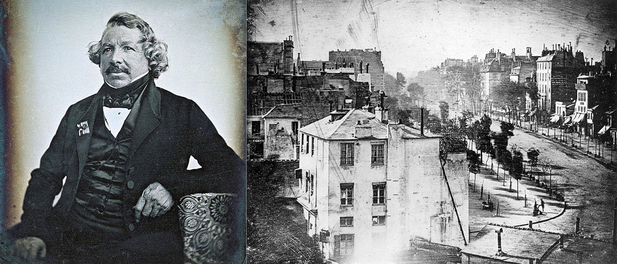
Within the museum’s walls, there are more than 400,000 photographic objects, including a collection of about 5000 daguerreotypes – images typically no bigger than a postcard, printed on polished copper plates and encased in glass. Sourced from all over the world, examples include a likeness of the “father” of the daguerreotype, French photographer Louis-Jacques-Mandé Daguerre. Invented by Daguerre in the 1830s, daguerreotypes became the first commercially available form of photography.
For about two decades – until other, more efficient photographic methods became available and popular – this early way of recording visual images surged in popularity worldwide. For the first time, people could buy an exact image of themselves. The method was used to capture snapshots of landmarks, famous people, ordinary citizens, as well as notable events such as burning buildings or lively street scenes. Subjects included Queen Victoria and her daughter, US president Abraham Lincoln, writer Harriet Beecher Stowe, the Acropolis in Athens and the waterfront of Cincinnati. The Eastman museum collection includes a daguerreotype of abolitionist Frederick Douglass.
“People went nuts with it,” says Patrick Ravines, a chemist-turned-conservator at Buffalo State College in New York, who studies and collects daguerreotypes. “They used to document everything around the world, and daguerreotypes show things that don’t exist any more.” He points out that researchers can study daguerreotypes to study erosion in natural settings, measure the growth and change of cities, or even see what archaeological sites – such as the Acropolis – looked like in the 19th century. These images serve as a record of their day.
Yet, like everything, daguerreotypes yield to time, and are now in trouble. Some have become cloudy; others have developed spots. Some are so damaged that they’re no longer recognizable. They’re deteriorating at a rapid clip.
In 2005 art conservator Ralph Wiegandt, then at the Eastman Museum, helped prepare a large exhibition of daguerreotypes to be exhibited at the International Center of Photography in New York City. During the exhibition, curators noticed that 30 of the objects showed signs of inexplicable degradation and were fading fast. To find ways to salvage the images, Wiegandt turned to physicists at the nearby University of Rochester.
One of them was Nicholas Bigelow, who was eager to bring his team to the project. Bigelow’s scientific interest was piqued by the fact that early photographers were using nanotechnology to create the images, even though they were unaware of it at the time. “If you were to take the nanoparticles that form the image on the daguerreotype, you’d have to have between 100 and 1000 of them stacked side by side to be as [wide] as a human hair,” he says in a video interview filmed for the University of Rochester.
To save the daguerreotypes, Bigelow and his fellow scientists would need to follow a three-step process. First, they’d have to understand the surface physics and chemistry of how the images were made. Second, they’d have to identify the processes that fuel degradation. Third, they’d have to get creative – and find ways to use their research to forestall the process, if not reverse it. Fortunately, the team had an arsenal of modern imaging tools at its disposal, including scanning electron microscopes (SEMs), transmission electron microscopes, X-ray scanners and focused ion beam milling, to get beneath the surface of the centuries-old images.
“It’s a surface where history and art and science intersect, and it’s very, very complicated,” says Brian McIntyre, who manages the Integrated Nanosystems Center at the University of Rochester. As he points out, the objects provide the first true – not drawn – images of the world. “They give us a window on the world that really is first light.”
Forming the image
Daguerreotypes aren’t like other photographs. The image is printed using a laborious process that may require hours just to get one shot. It’s printed directly onto a metal plate – which renders each daguerreotype one-of-a-kind and irreproducible. They’re unusually rich in detail. Bigelow says he was drawn to the project after seeing a daguerreotype of the Cincinnati waterfront made in 1848. Zooming in on that image with magnifying lenses reveals an astonishing level of detail. “If you blow this up, and you blow it up, and you blow it up, the level of resolution and detail in this nanotechnology photograph is fantastic,” he says.
Wiegandt, who now continues his daguerreotype work as a visiting research scientist at the University of Rochester, says that well-made daguerreotypes can be enlarged 20–30 times and still reveal minute details of their subjects – a resolution that, today, would require a 140,000-megapixel digital camera.
But the scientists wanted to zoom in even further to see what was happening at the nano level. “It was determined that looking at the surface – the surface science, the surface morphology, the surface physics – was where we ought to start,” recalls McIntyre.
The process of making a daguerreotype begins when a silver-coated copper plate is painstakingly polished to a mirror finish. The plate is then placed in a closed box with iodine and bromine vapours, which sensitizes it to light and gives it a rose-coloured hue. Next, the picture is taken: the plate is mounted in a camera and exposed to light, usually for a few minutes, to form the image. The plate is then exposed to hot mercury vapours, which create particles of amalgam – a combination of silver and mercury – that develop the image. Finally, the silver iodide is removed, and the image may be “gilded”, which involves coating it with gold chloride.
“The fascinating aspect of daguerreotypes is that you’ve got light interacting with a material – and changing the nature of that material,” says Ravines, who led a study, published in May in the Journal of Imaging Science and Technology (60 30504), that reports what happens at the molecular level during that laborious process. He and his colleagues focused primarily on the steps during which the image was formed – after the plate was exposed to iodine and bromine, but before it was gilded.
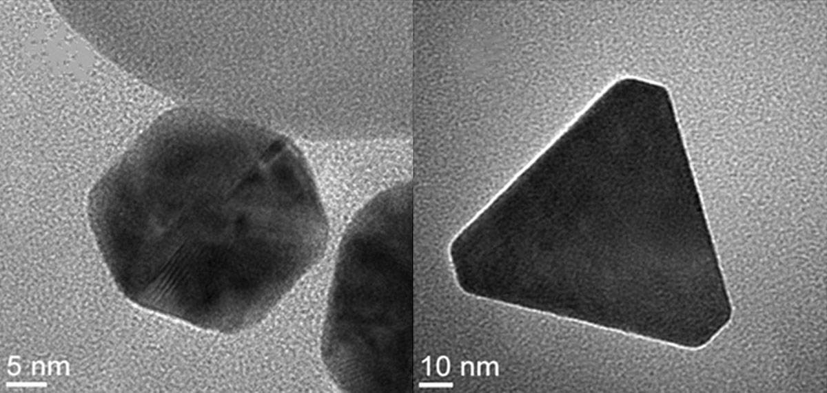
Images taken with an SEM of a daguerreotype from Ravines’ personal collection showed that many of the larger amalgam particles – about 200–800 nm in diameter – are hexagonal in shape and pile up in the whites and light greys of the image (figure 1). Their findings corroborate studies by scientist Alice Swan in the late 1970s, and by art conservation scientist M Susan Barger in the 1980s and 1990s, that also revealed the hexagonal shape. In the new study, however, the researchers observed particles with other shapes too, including large trapezoidal solids and other 3D geometries. Ravines suspects those other geometries may correspond to a variety of amalgam species that combine mercury and silver in different proportions.
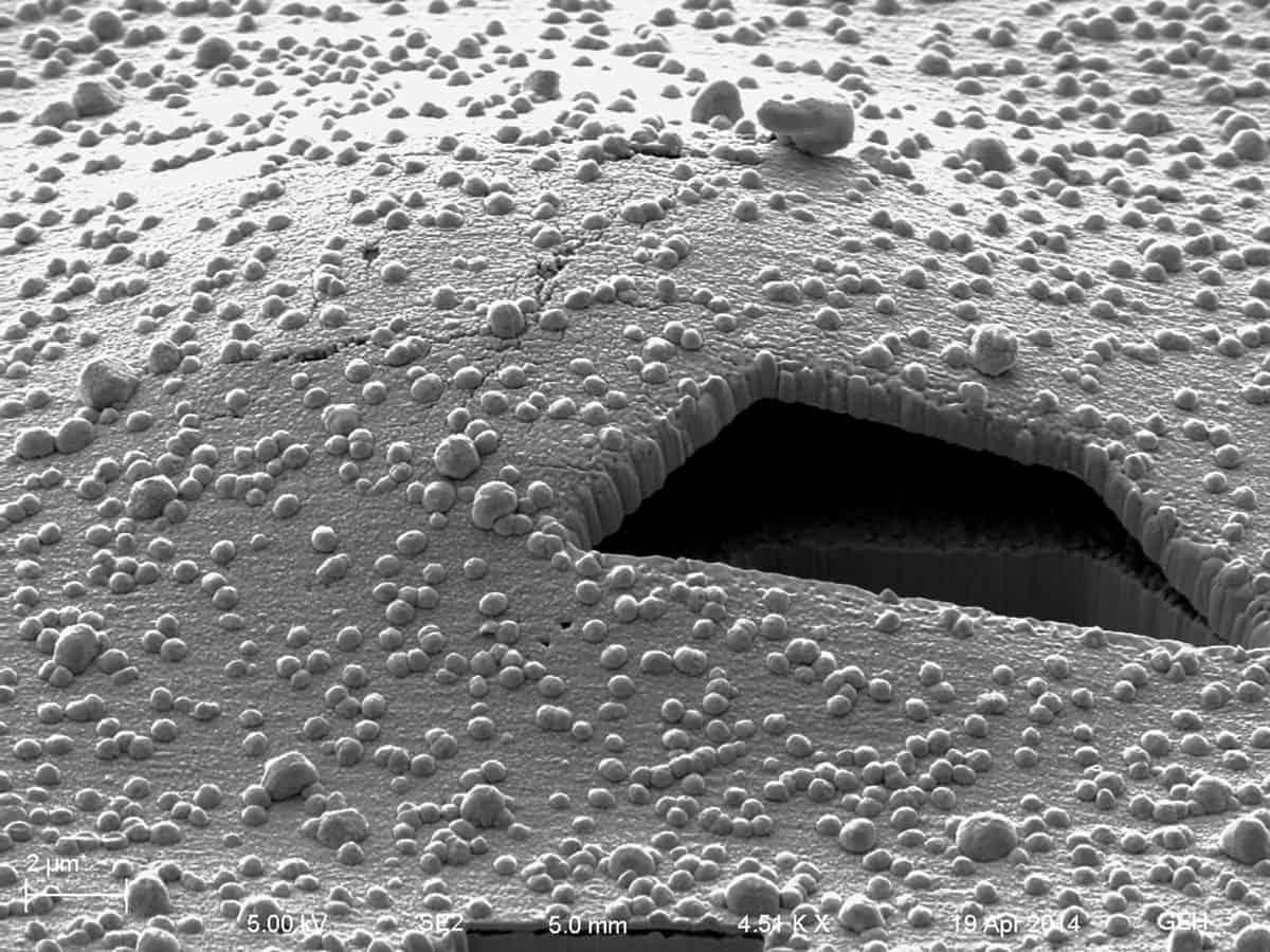
Understanding how nanoparticles and the surface respond to airborne pollutants such as water vapour, sulphur-based gaseous pollutants and other sources of corrosion, says Ravines, could help scientists better observe how the images vanish over time. Figure 2 shows one example of the damage.
Destructive forces
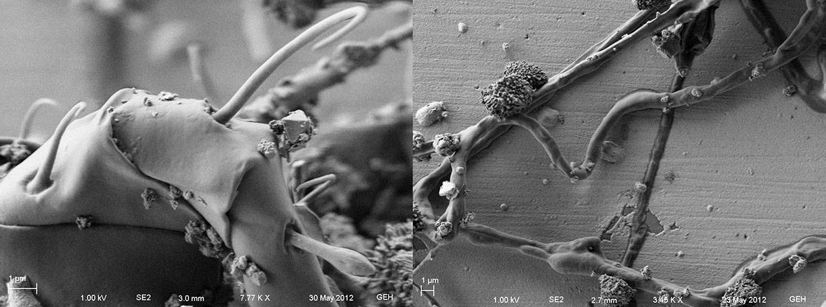
In 2010, with support from a grant from the National Science Foundation, Bigelow, Wiegandt and McIntyre went looking at damage on the nanoscale. They were in for some surprises. Perhaps most startling was the fact that the daguerreotype surface can support life that, ultimately, harms it. “One thing that we never would have expected is that the daguerreotype is a biologically active surface,” says Bigelow. “We have discovered essentially on every daguerreotype we looked at, there are small colonies of fungi growing, and those fungi are damaging the surface.”
SEM images reveal networks of micron-scale filaments sprawling on the surface (figure 3). The microbes seemed to not only live on the surface, but also interact with it in some way – a discovery that McIntyre likens to finding life on Mars. The surface of a daguerreotype is “pretty darn toxic”, as he puts it. Silver and mercury have antimicrobial properties, but they don’t stop the micro-organisms from spreading across the silver surface of the images.
The team also found problems lurking beneath the surface. “One thing we’ve observed is that below the image particles, and above the native silver surface, there seems to be a gap forming, and that gap can lead to the uppermost surface of the daguerreotype exfoliating from the underlying silver,” says McIntyre. In extreme cases, this condition leads to large flakes peeling away from the image like ripped pieces of paper – a type of degradation that’s easily visible to the naked eye. In images from SEMs, the gaps resemble networks of underground caves, with the scientists saying these look like Kirkendall voids – cavities that appear at the interface between two attached metals, such as a metal and an alloy in a solder joint. In daguerreotypes, the voids likely form as a result of the reaction between a gold (gilding) solution and the silver. Though the exfoliation mechanism isn’t well understood, those SEM images suggest that the metal seems to be moving, in part, due to it interacting with the solution used to gild the image, though McIntyre admits other factors could be involved.
Writing in the Journal of Imaging Science and Technology, Ravines says he has seen voids beneath the image that raise questions about the overall stability of the surface that the image rests on. Understanding those voids will be important for devising sound conservation treatment strategies. “We have to be careful because of the way that subsurface is almost like Swiss cheese,” he says. “Knowing the way [the daguerreotype] is built allows us to better preserve it.”
Another source of degradation comes, ironically, from historic efforts to preserve the images. Daguerreotypes are typically sealed in cases beneath a cover glass. Over time, moisture accumulates on the underside of the cover glass and dissolves it. The highly alkaline droplets can drip onto the daguerreotype. “We see patterns of degradation associated with the glass coming apart,” McIntyre says. “You try to do the right thing 150 years ago, but your best intensions are foiled by nature.”
Rescue strategy
The first priority in conserving daguerreotypes is to stabilize them in their current state. Following on from the imaging work, Wiegandt began to develop new strategies to protect the artefacts from degrading any further. One tactic included encapsulating the image in a frame filled with an inert gas, such as argon, to prevent pollutants and other corrosive elements from getting inside. That may not be a permanent solution, but it might buy more time. “If you can give the daguerreotype 20 years of life before a recharge, that’s a good investment of time and energy,” says Ravines.
Another tantalizing possibility, says McIntyre, is to undo the damage altogether. “We’ve done some things at the nano level that we think can not only stabilize but perhaps reverse some of the degradation process,” he says. One approach addresses the surface-exfoliation problem In preliminary experiments, he says, the researchers have been developing a way to physically repair that damage – a bit like a nano sewing machine that might stitch the damaged area.
McIntyre cautions, however, that so far they’ve only been able to use this approach in micron-sized areas, and scaling it up to be useful on daguerreotypes in general would be difficult. “I don’t know, frankly, if it’s possible, but it’s certainly conceivable,” he says. “It would be very cool.” Developing a useful tool would also not come cheap.
Daguerreotypes today
Daguerreotypes, in a way, have now come back into fashion. Professional photographers and curious hobbyists can learn how to make them in classes at the Eastman Museum, and successful artists including Adam Fuss have created oversized daguerreotypes many times larger than anything produced in the 19th century. In another 100 years, those works will likely face degradation, too – though perhaps, by then, conservators will have a set of sophisticated tools to keep the images from fading away.
But the benefits of saving daguerreotypes may even extend beyond the art world, with Bigelow and Wiegandt hoping their findings will have applications to other areas of nanotechnology. The amalgam nanoparticles, for example, exhibit “self-assembling” behaviour – a growing area of nanoscience research.
For McIntyre, the project not only demonstrated the interdisciplinary reach of nanoscale physics – but was a professional thrill too. “It’s probably one of the most interesting things I’ve ever worked on,” he says. “It’s not often you get the opportunity to work in the historical realm, the science realm and the art realm simultaneously.”