Since the inception of quantum mechanics, physicists have sought to understand its repercussions for our universe. One of the theory’s stranger consequences is entanglement: the phenomenon whereby a pair or group of particles becomes connected in such a manner that the state of any one particle cannot be described independently. Instead, its state is intrinsically correlated with the state of the other(s), even if the particles are separated by large distances. As a result, a measurement performed on a particle in an isolated location can affect the state of its entangled twin far away.
Researchers at the California Institute of Technology (Caltech) in the US have now discovered a way to use this quantum property to double the resolution of optical microscopes. The new technique, dubbed quantum microscopy by coincidence (QMC), illustrates the advantage of quantum microscopes over classical ones, and could have applications in non-destructive imaging of biological systems such as cancer cells.
Quantum microscopy by coincidence
An optical (light) microscope can resolve structures that are about half the wavelength of the light used. Anything smaller than that cannot be distinguished. Therefore, a possible route to improved resolution is to use higher intensities and shorter wavelengths of light.
But there is a caveat. Shorter wavelengths of light have higher energies, and this highly energetic light can damage the object being imaged. Living cells and other organic materials are particularly fragile.
In the latest work, which appears in Nature Communications, a team led by Lihong Wang used a pair of entangled photons, or biphotons, to circumvent this roadblock. The photons that make up the biphoton pair do not have an individual identity and they necessarily behave as a composite system. But, crucially, the wavelength of these composite photons is half the wavelength of an unentangled, classical photon at the same energy. Therefore, a biphoton pair carrying the same amount of energy as a classical photon can achieve double the resolution.

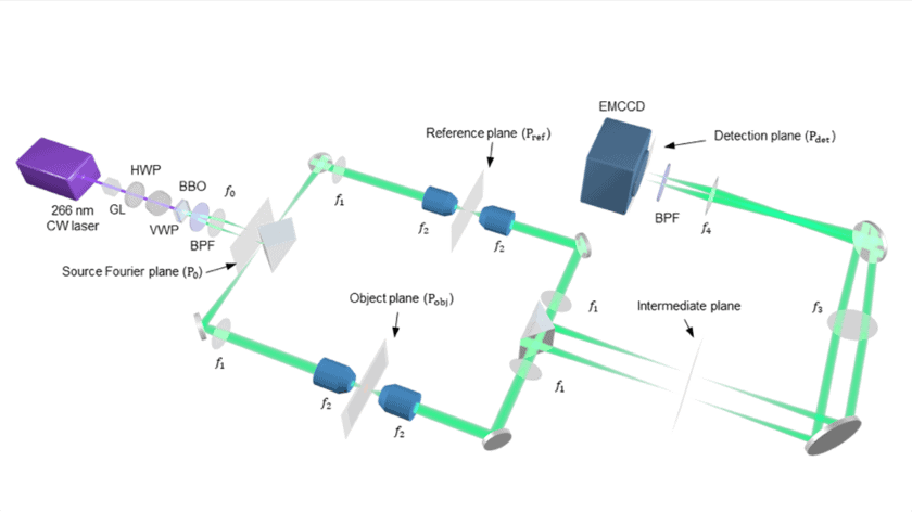
To demonstrate this, Wang and colleagues used a crystal to split an incoming photon into an entangled biphoton pair made up of a signal photon and an idler photon. These biphotons travel along symmetric paths designed using a network of mirrors, lenses and prisms. The signal photon traverses the path containing the object being imaged, whereas the idler photon travels unobstructed. Eventually, both photons reach a detector plate, which records the information carried by the signal photon. This information is then correlated with the detection of the idler photon’s state and used to create an image.
Advantages over classical microscopy
The concept of using entangled photons to enhance imaging is not new, but it has previously been limited to imaging larger objects. The Caltech team is the first to demonstrate a viable setup that can resolve details down to the cellular scale. Using the spatial and temporal correlations between the signal and idler photon measurements (which do not exist for classical photons), Wang and colleagues also showed that the QMC method has advantages over classical microscopy in terms of noise resistance and image contrast.

So far, the team has demonstrated the advantages of QMC through the bioimaging of cancer cell (see photo above). According to Wang, other applications could include non-destructive imaging of photosensitive materials such as organic molecules and memory devices. Additionally, since QMC produces a twofold improvement in the resolution of the microscope, any future advances in classical microscopy could be further enhanced by leveraging this property of quantum microscopy.
Quantum microscope uses entanglement to reveal biological structures
But while QMC has much promise, a major challenge when compared with state-of-art classical microscopes is speed. Current methods to create entangled photons are inefficient, resulting in a low output of biphoton pairs. Since any advantage of QMC relies on being able to generate an abundance of biphotons, developing methods that can accomplish this will be crucial. “The development of strong and/or parallel quantum sources for quantum imaging is expected to speed up data acquisition,” Wang tells Physics World. Once that happens, quantum imaging techniques will truly come to the forefront of microscopy.


