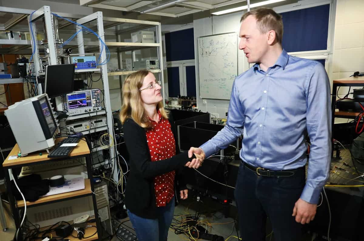
A novel detection method for optical coherence tomography (OCT) achieves high-quality imaging at low light intensity. The new OCT method was developed by a group of researchers from the University of Auckland in New Zealand and the Nicolaus Copernicus University in Poland. Borrowing ideas for their detection mechanism from quantum optics, they describe in Optics Letters how the method can produce comparable results to standard OCT systems at very low light intensity levels (around 10 pW).
OCT is a widely used medical imaging technique that can non-invasively obtain cross-sectional images of semi-translucent materials. It has applications in several medical disciplines, including ophthalmology, cardiology and dermatology, where its resolution allows visualization of microscopic details. It works by illuminating the tissue with broad-bandwidth visible or near-infrared light. This light is scattered and reflected by the biological media and then captured and assessed using a spectroscopic detector.
OCT can provide images of tissue morphology at 1 µm resolution and it is now a common part of routine eye exams, where an ophthalmologist may recommend the scan to inspect the retina’s distinctive layers and rule out glaucoma or retinal diseases.
In a clinical setting, OCT is limited by the allowed intensity levels. For example, the imposed safety levels for imaging the eye are 1.7 and 5 mW, at wavelengths of 800 and 1060 nm, respectively, constraints that can sometimes affect the fidelity of the obtained images. A team led by Sylwia Kolenderska bypassed this limitation by using a superconducting single-photon detector (SSPD) instead of the standard OCT detector – an idea inspired by quantum optics, where SSPDs are used to study various properties of single photons.
One photon at a time…
Leading the way towards high-fidelity low-power OCT systems, the team came up with the idea of using an SSPD while researching new OCT detection schemes. As these sensors can detect single photons, the proposed setup uses a much smaller amount of light than a standard OCT system. In fact, the required power is a few orders of magnitude smaller than clinically available counterparts.
To make the new detection scheme work, the team had to make a few changes to the original optical setup, which requires a way of discerning the different wavelengths of light being reflected from the imaged object. As they were now working in a single-photon detection regime, the researchers decided to couple the SSPD with a long (5 km) fibre spool that induces a wavelength-dependent time delay. In other words, different wavelengths of light will travel at different speeds down this fibre, enabling the SSPD to capture the light spectrum.
…yields high-quality images
The team tested the new OCT setup using a 1550 nm pulsed light source to image two objects: a stack of different types of glass and a slice of onion. The glass stack was made up of three layers: quartz (50 µm thick), sapphire (460 µm thick) and BK7 glass (500 µm thick), and it was used as a controlled test to understand how well the different layers can be discerned. The slice of onion was used as a biological sample. Both experiments resulted in good-quality images, comparable with images obtained by standard OCT systems, but using intensity levels five orders of magnitude lower.
The researchers point out that they observed artefacts in the reconstructed images. These are unwanted image features that, in this case, appear because the detection system is recording all types of interactions between photons inside the sample. Artefact removal algorithms can be used to clean up the images and are straightforward to use when the imaged object has a well-defined structure, such as the stack of glass. These artefacts become more problematic when dealing with biological samples, where the structure is highly heterogeneous and complicated.


