A research paper describing a texture-based model that predicts lung metastasis risk in soft-tissue sarcomas has won its authors the 2018 Physics in Medicine & Biology (PMB) citations prize. This annual prize recognizes the PMB paper that received the most citations in the preceding five years.
The paper, A radiomics model from joint FDG-PET and MRI texture features for the prediction of lung metastases in soft-tissue sarcomas of the extremities, was written by researchers from McGill University. The authors describe the development and evaluation of a model that analyses FDG-PET and MR images using radiomics — in which large amounts of quantitative features are extracted from medical image data.
“Quantitative information from imaging has been underutilized, with simple (first order) features unable to provide necessary diagnostic or predictive guidance, which radiomics (higher order features) is able to compensate for,” says senior author Issam El Naqa. “Multi-modality imaging can provide a better representation than any single modality. We aimed to develop a robust approach for radiomics while avoiding overfitting with large number of variables, which is a common problem in the field.”
About one quarter of patients with soft-tissue sarcomas develop distant metastases, rising to roughly half for those with high-grade tumours, with the lungs the main site of metastases. A model that can assess lung metastasis risk at the time of diagnosis could enable better adapted treatments and improve survival.
In many solid tumours, heterogeneity is associated with higher metastasis risk, thus the researchers employed texture analysis to quantify tumour heterogeneity. They retrospectively evaluated FDG-PET and MRI scans from 51 patients with soft-tissue sarcomas, extracting features from the tumour region of separate and fused scans. “The idea is that with radiomics, the spatial distribution of image intensities can reveal tumour aggressiveness, allowing for early intervention,” explains El Naqa.
They found that texture features extracted from fused PET/MRI scans significantly outperformed those from separate scans in terms of lung metastases prediction. The researchers identified a combination of four texture features that delivered the best predictive performance and used these to create the predictive model.
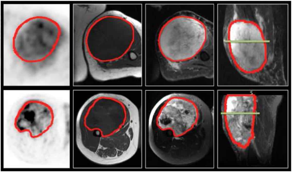
“We observed that patients who developed lung metastases often exhibited large low-uptake regions in the inner portion of their tumour on FDG-PET scans, most likely representing necrotic areas,” explains first author Martin Vallières. “The presence of such regions suggests that the tumour is rapidly increasing in size and might be more at risk to metastasize. The model we developed contains texture metrics that most likely detect the presence of these sub-regions inside other regions, for example, high-uptake tumour sub-regions.”
Moving ahead
The paper’s high number of citations is likely due to its introduction of new methods for combining multi-imaging modalities and applying texture analysis to fused hybrid PET/MR images. The authors also developed texture optimization techniques to enhance predictive values and created a robust strategy for constructing radiomics-based prediction models. In addition, notes Vallières, all of the imaging data and code are shared online (on The Cancer Imaging Archive and GitHub), allowing others to fully reproduce the results.
Since the paper was published, the researchers have tested their prediction model on a prospective cohort of patients. They found that the model provided comparable performance to original estimates, when uncertainties were accounted for (phiRO 6 53).
“In that work, we also verified a strategy for soft-tissue sarcoma management,” notes Vallières. “This involves: identifying patients at higher risk of developing lung metastases using the developed radiomics-based prediction model; delineating hypermetabolic and hypoxic tumour sub-volumes via functional imaging; and planning radiotherapy delivery with dose escalation to tumour sub-volumes.”
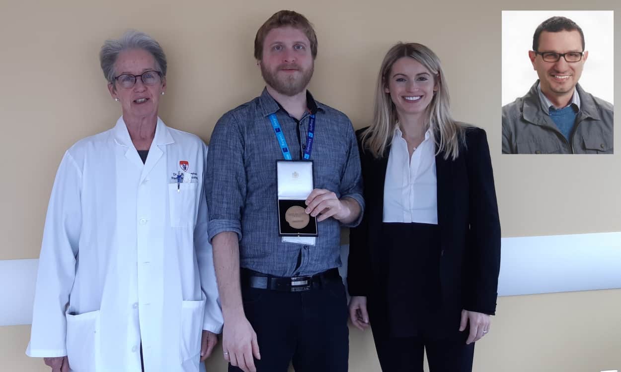
The PMB citations prize is marked with the presentation of the Rotblat medal, named in honour of Sir Joseph Rotblat, PMB’s second and longest-serving editor. “I feel honoured to receive this prize,” El Naqa tells Physics World. “I agree with Issam, this is an honour!” adds Vallières.
• The winner of the 2018 Physics in Medicine & Biology citations prize is: A radiomics model from joint FDG-PET and MRI texture features for the prediction of lung metastases in soft-tissue sarcomas of the extremities M Vallières, C R Freeman, S R Skamene and I El Naqa Phys. Med. Biol. 60 5471


