ESTRO 2023, the annual meeting of European Society for Radiotherapy and Oncology, saw over 8000 delegates head to Vienna last week. And judging by the crowds on the exhibit floor, all were keen to check out the latest product developments, hear research updates at the exhibitor’s booths, or just join the long queue for GE Healthcare’s ice cream. Here are just a few of the products that caught my eye at this year’s trade show.
Heating up treatments
Hyperthermia, heating tumours to around 41–43°C, can enhance the effects of both chemo- and radiotherapy. Italian manufacturer Med-Logix has developed a dedicated deep hyperthermia system – the ALBA 4D – that uses a multibeam phased array of four waveguide antennas to precisely focus radiofrequency fields onto the tumour to raise its temperature.
The ALBA 4D can focus energy onto targets at any depth and location in the pelvis, abdomen and extremities. The system, which includes a robotic gantry for precise patient positioning, automatically tracks the temperature and location of the focal zone, heating the target in 5–10 min while avoiding overheating of healthy tissues.
Hyperthermia impacts radiotherapy via three mechanisms: inhibiting repair of DNA damage; reoxygenation via increased tissue perfusion; and direct cell killing. These effects can be used to create a higher equivalent radiation dose, or alternatively, to deliver a lower dose while maintaining the same tumour killing effect.
For use with radiotherapy, Med-Logix’s Sara Baghaei explained, the hyperthermia should be delivered within one hour of the radiation treatment. The ALBA system can also be employed to enhance chemotherapy, where it can be used simultaneously with drug delivery.
“The technology allows users to perform fast hyperthermia with high temperatures,” said Baghaei. “It’s like we are giving a higher dose of radiation without any extra toxicity.”
MRI coils made for radiotherapy
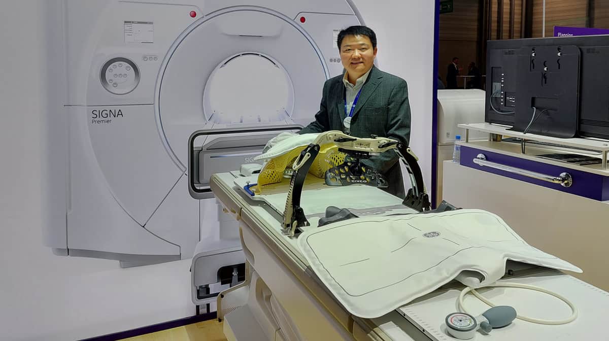
With researchers investigating the feasibility of MRI-only radiotherapy planning, GE Healthcare highlighted its AIR Coils – the first MRI coils designed specifically for radiation oncology patient scans. Available for brain, head-and-neck or body imaging, the AIR Coils are light, flexible and comfortable for the patient.

According to GE HealthCare’s global product manager Michael Mian, one big advantage of the AIR Coils is that they do not require supports, which are usually placed between rigid MRI coils and the patient to avoid anatomy distortion during imaging.
“This opens up space for patient immobilization devices,” Mian explained. It also means that the coils can be placed closer to the patient, thereby improving the quality of the image. “For the first time, you can use the coil suite to deliver diagnostic image quality with the patient in the immobilization device,” he said.
The AIR Coils are also easy are to use, an important factor when bringing together radiology and radiation oncology departments that may be used to different patient set-up procedures. “The coil design is so simple, you don’t require extensive training to learn how to use it,” said Mian.
Enhancing target visibility
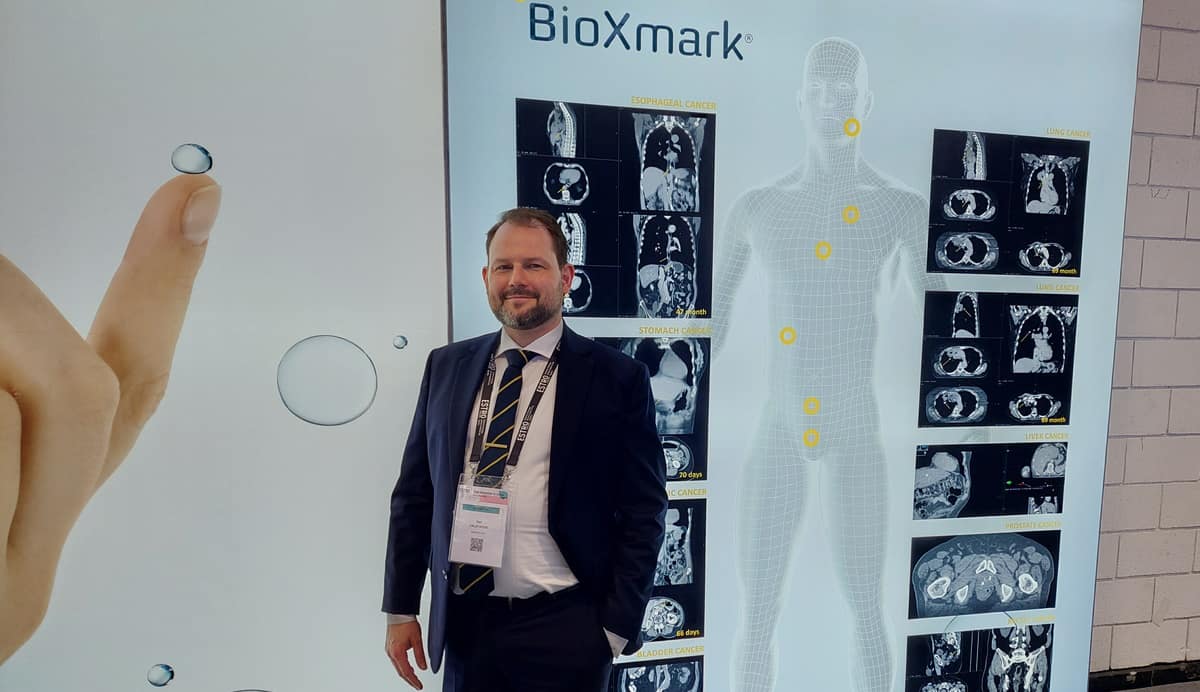
Danish medical device company Nanovi showcased BioXmark, its unique liquid fiducial marker. Fiducial markers, used as target reference points to guide radiation therapy and increase treatment accuracy, usually consist of small metal implants. BioXmark is different, consisting of a biocompatible long-chain carbohydrate containing iodine for contrast.
To create the fiducial marker, a small volume (around 80 µl) of BioXmark is injected into the body, where it changes viscosity from a liquid into a consistency similar to chewing gum. This soft marker can then be visualized on X-ray, CT or MRI scans for treatment planning, radiotherapy guidance or follow-up.
Once in place, the fiducial is highly stable, with studies revealing that it is still visible up to 69 months after implantation. “We have not seen it disappear yet,” said Nanovi’s Dan Calatayud. He noted that the liquid fiducial is easier to implant than metal markers, and that the team had demonstrated “significantly shorter implantation times”.

The non-metallic composition of BioXmark leads to a low level of artefacts on X-ray based imaging; it also offers low dose perturbation when used with proton therapy. But BioXmark’s main advantage, said Calatayud, is that it can be used within thin-walled, hollow organs – such as the oesophagus, stomach and bladder, for example – where it is extremely difficult to implant a piece of metal. Currently, fiducial markers are only established in prostate and breast treatments.
“We want to open up the possibility of taking this precision into new indications,” he explained.
Surface-guided radiation therapy
Brainlab unveiled its ExacTrac Dynamic Surface system for radiotherapy patient positioning and monitoring. Surface-guided radiotherapy enables precise tracking of the patient’s surface and breathing motion. This in turn allows gating of the radiation beam so that the tumour is only irradiated when in the planned position.
The system is based around two 3D cameras housed in a centralized camera unit, which emit a structured blue light pattern on the patient surface. The cameras project 300,000 points onto the patient, these are then matched to a heat signal obtained by thermal camera within the same unit.
A stroll around the ESTRO show floor
“The thermal signal gives an additional fourth dimension for extra precision,” explained Brainlab’s Carsten Sommerfeldt. He noted that with a three-camera positioning system, a moving gantry can often block one of the cameras. The single unit, however, is always in the line-of-sight to the patient and can constantly track the patient’s surface during beam delivery.
ExacTrac Dynamic Surface is designed to operate with Varian’s Edge and TrueBeam radiotherapy systems, as well as Elekta’s Versa HD.


