Abnormalities with the nervous system can cause an overactive urinary sphincter, leading to urinary incontinence. Researchers from University College London (UCL) and Nephro-Urology Clinical Trials (NUCT) Ltd have designed an ano-rectal surface electromyography (sEMG) probe to indirectly monitor muscle activity of the external urethral sphincter. The estimated amplitude of this muscle activity could be used as a threshold to initiate trans-rectal stimulation to treat overactive bladders (Physiol. Meas. 38L17).
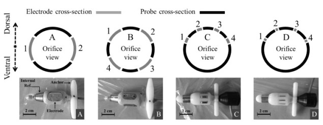
Research suggests that an overactive urethral sphincter muscle could be identified by monitoring the activity of the external anal sphincter (EAS) muscle. UCL’s Arsam Shiraz, under the leadership of Andreas Demosthenous, has worked with a group of researchers to determine a feasible sEMG probe design to detect such signals. The researchers manufactured four probes with a diameter of 18 mm, and with electrode configurations that varied in the number of electrodes, their size and spacing.
Proof of concept
To attain an acceptable design for an intra-anal sEMG probe, the team tested the four probes on a healthy volunteer by inserting each probe through the anal orifice. The individual was required to contract their EAS muscle three times, lasting 10 seconds each time.
The researchers used correlations between the time of EAS contractions and the sEMG traces to determine a viable in situ probe design. In some probes, they observed poor quality signals and odd findings – such as moments of silence during EAS contractions – which they assumed were related to the large electrode sizes, unstable electrode-tissue contact and the placement of the reference electrode. However, they successfully designed one probe – probe C – which produced the signal that was most correlated with EAS muscular activity.
The researchers had specific design constraints when developing the prototype, such as patient comfort due to the long periods of time that the probe would be inserted and maintaining good contact with the ano-rectum. The final prototype was based on design C with electrode dimensions of 3 mm wide and 1 cm long, and an electrode spacing of 5 mm.
Potential future applications of the probe include the embedding of stimulator electrodes to allow nerve stimulation for the treatment of overactive bladders. By recording intra-anally for a period of time, the EAS sEMG signal could provide a trigger for stimulation by the electrodes.


