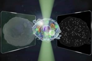
Physicists in Japan have developed a new technique for determining the identity of groups of individual atoms. The technique is an improvement on existing characterization methods in microscopy, which can only detect the positions of atoms and not their specific chemical type. It has been used by the researchers to detect the composition of a substance to a resolution of 10 nm.
Dubbed Synchrotron Radiation Scanning Tunnelling Microscopy (SRSTM), the new technique has been developed by Taichi Okuda and colleagues at the Institute and the University of Hyogo, in Japan. It involves placing a sample of interest in the intense x ray beam of a synchrotron source.
Photons from the beam excite core electrons in the sample’s atoms, which then spit out “secondary” electrons as they decay back down to their ground states. These secondary electrons are then detected by the tip of the scanning tunnelling microscope (STM) as they tunnel across the gap. The size of the current depends on the specific type of atom that has produced the secondary electrons, which means that each element has a unique “fingerprint” (Phys. Rev. Lett. 102.105503).
The Japan team tested its technique on a checker board-patterned sample of nickel and iron on a gold film. Each square of nickel and iron was one micron wide and 10 nanometres thick. Using the BL-13C beam line at the KEK Photon Factory in Japan, they found they could distinguish the individual elements with a spatial resolution of just 10 nm.
“We are now trying to further improve the spatial resolution and signal-to-noise ratio in our technique,” Okuda told physicsworld.com. “We will also apply the method to magnetic materials and observe magnetic domain structures,” he added.


