An alarm goes off, shocking you into consciousness. You blearily mute the too-loud beeping and try to reacquaint yourself with reality, lumbering out of bed. It takes about 30 minutes before the grogginess has dissipated…
Waking up can feel like an emergency stop on our nightly ruminations; the slow return to some semblance of normality can take a few minutes or many more, depending on whether one is sleep deprived, and in which sleep cycle one wakes from. Fast recovery from this leftover “inertia” is crucial for those operating in emergency conditions, such as medical or military staff, for example, and hence a deeper understanding of this concept would be valuable.
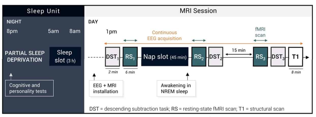
Scientists at the Lyon Neuroscience Research Center in France have probed the cerebral correlates of sleep inertia using a combination of electroencephalography (EEG), functional MRI (fMRI) and a behavioural task. Combining multiple resting state fMRI scans and continuous EEG allowed the researchers to measure brain function just before a 45-min afternoon nap, 5 min immediately after awakening and then 25 min after.
Post-awakening, participants had fewer responses in the behavioural task, increased EEG delta-band power (brain waves oscillating at 0.5-4.5 Hz), and reduced anti-correlation (as usually seen when awake) between brain networks (NeuroImage 10.1016/j.neuroimage.2018.09.033).
Measuring sleep inertia
After a night of only three hours sleep, 34 participants were hooked up to a polysomnographic cap (an EEG cap with nine electrodes) to measure the tiny summed electric fields of thousands of neurons. Over time, these brain waves can be grouped into different frequency bands, for example, delta waves at 0.5-4.5 Hz.

Next, the subjects performed a descending subtraction task (DST) in which they had to subtract a suite of preceding numbers from a given a three-digit number. This was done prior to a 6 min resting-state fMRI scan. Following this, participants had the 45 min afternoon nap (monitored continuously by EEG) and were then awakened: 14 during N2 sleep and 20 during N3. Five minutes after awakening, a second resting state fMRI scan was acquired, followed by a repeat of the DST; 25 min after awakening, the fMRI and DST were repeated again.
The researchers extracted sleep statistics from the EEG hypnogram, such as which sleep cycles were attained and sleep efficiency. They also performed functional connectivity analysis on the fMRI data, correlating mean, spontaneous BOLD (blood oxygenation level dependent) fluctuations from predefined network regions of interest (ROIs) to mean BOLD time series from every other ROI (within and between networks).
Sleep intrudes into wakefulness
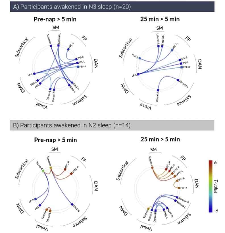
The authors suggest that increased EEG delta power at 5 min compared with 25 min post-awakening could be a signature of sleep inertia. Additionally, a reduced anti-correlation between two different default mode networks measured with fMRI (as compared to waking) at 5 min compared with 25 min post-awakening, is indicative of certain sleep-specific features “intruding” into wakefulness.
The lack of difference within and between networks pre-afternoon nap compared with 25 min post-awakening from the nap implies that sleep inertia has dissipated by this time, as evidenced by the connectivity returning to “normal”.
Although there was little difference in the EEG or behavioural results based on whether participants were awakened from N2 or N3 stages, the researchers noted a larger disruption in functional connectivity from N3-to-awake than N2-to-awake.
From N2-to-awake, the biggest differences in ROI-to-ROI connectivity were seen when comparing 25 min to 5 min post-awakening, whereas from N3-to-awake, it was when comparing pre-afternoon nap to 5 min post nap. This suggests that the dissipation of sleep inertia is “faster” when waking from N2, than N3.
Feeling awake yet?
This study shows that sleep-specific brain activity does not disappear immediately after we wake up, but rather, this inertia persists for several minutes post-awakening. This phenomenon is associated with a decrease in cognitive performance, an increase in delta power and a disruption of functional networks, with this loss of functional segregation (how separate brain networks appear to be) worse when waking from deeper sleep (N3 versus N2). This exciting work paves the way for possible extension to studying sleep in more depth, for example measuring sleep inertia following N1 and REM sleep.