Portable multiphoton microscopy system uses femtosecond laser pulses to create pathology-quality diagnostic images

Multiphoton microscopy is a nonlinear optical imaging technique that enables label-free, damage-free biological imaging. Performed using femtosecond laser pulses to generate two- and three-photon processes, multiphoton imaging techniques could prove invaluable for rapid cancer diagnosis or personalized medicine.
Imaging biological samples with traditional confocal microscopy requires sample slicing and staining to create contrast in the tissue. The nonlinear mechanisms generated by femtosecond laser pulses, however, eliminate the need for labelling or sample preparation, revealing molecular and structural details within tissue and cells while leaving the sample intact.
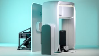
Looking to bring these benefits to cancer diagnostics, Netherlands-based start-up Flash Pathology is developing a compact, portable multiphoton microscope that creates pathology-quality images in real time, without the need for sample fixation or staining.
Fast and accurate cancer diagnosis with higher harmonic imaging
The inspiration for Flash Pathology came from Marloes Groot of Vrije Universiteit (VU) Amsterdam. While studying multiphoton microscopy of brain tumours, Groot recognized the need for a portable microscope for clinical settings. “This is a really powerful technique,” she says. “I was working on my large laboratory setup and I thought if we start a company, we could transform this into a mobile device.”
Groot teamed up with Frank van Mourik, now Flash Pathology’s CTO, to shrink the imaging device into a compact 60 x 80 x 115 cm system. “Frank made a device that can be transported, in a truck, wheeled through corridors, and still when you plug it in, it’s on and it produces images – for a nonlinear microscope, this is pretty special,” she explains.
Van Mourik has now built several multiphoton microscopes for Flash Pathology, with one of the main achievements the ability to measure samples with extremely low power levels. “When I started in my lab, I used 200 mW of power, but we’ve been able to reduce that to 5 mW,” Groot notes. “We have performed extensive studies to show that our imaging does not affect the tissue.”
Flash Pathology’s multiphoton microscope is designed to provide rapid on-site histologic feedback on excised tissue, such as diagnostic biopsies or tissue from surgical resections. One key application is lung cancer diagnosis, where there is a clinical need for rapid intraoperative feedback on biopsies. The standard histopathological analysis requires extensive sample preparation and can take several days to provide results.

“With lung biopsies, it’s challenging to obtain good diagnostic material,” explains Sylvia Spies, a PhD student at VU Amsterdam. “The lesions can be quite small and it can be difficult to get to the right position and take a good sample, so they use several techniques (fluoroscopy/CT or ultrasound) to find the right position and take multiple biopsies from the lesion. Despite these techniques, the diagnostic yield is still around 70%, so 30% of cases still don’t get a diagnosis and patients might have to come back for a repeat biopsy procedure.”
Multiphoton imaging, on the other hand, can rapidly visualize unprocessed tissue samples, enabling diagnosis in situ. A recent study using Flash Pathology’s microscope to analyse lung biopsies demonstrated that it could image a biopsy sample and provide feedback just 6 min after excision, with an accuracy of 87% – enabling immediate decisions as to whether a further biopsy is required.
“Also, many clinical fields are now focusing on a one-stop-shop with diagnosis and treatment in one procedure,” adds Spies. “Here, you really need a technique that can rapidly determine whether a lesion is benign or malignant.”
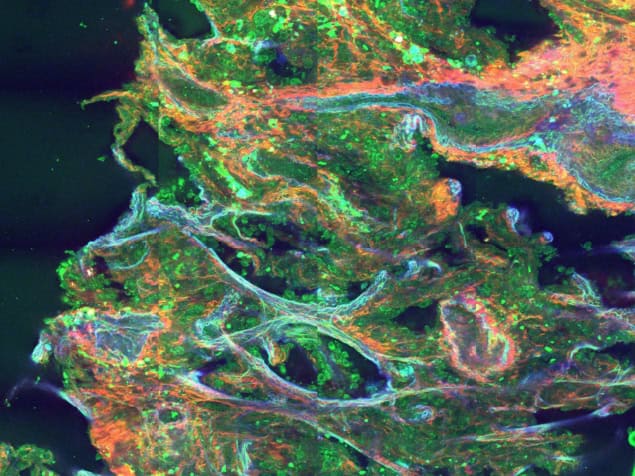
The microscope’s impressive diagnostic performance is partly due to its ability to generate four nonlinear signals simultaneously using a single ultrafast femtosecond laser: second- and third-harmonic generation plus two- and three-photon fluorescence. The system then uses filters to spectrally separate these signals, which provide complementary diagnostic information. Second-harmonic generation, for example, is sensitive to non-centrosymmetric structures such as collagen, while third-harmonic generation only occurs at interfaces with differing refractive indices, such as cell membranes or boundaries between the nucleus and cytoplasm.
“What I like about this technique is that you can see similar features as in conventional histology,” says Spies. “You can see structures such as collagen fibres, elastin fibres and cellular patterns, but also cellular details such as the cytoplasm, the nucleus (and its size), nucleoli and cilia. All these tiny details are the same features that the pathologists look at in conventional histology.”
Applying femtosecond lasers for 3D-in-depth visualization
The femtosecond laser plays a key role in enabling multiphoton microscopy. To excite two- and three-photon processes, you need to have two or more photons in the same place at exactly the same time. And the likelihood of this happening increases rapidly when using ultrashort laser pulses.
“The shorter the pulses are in the time domain, the higher the probability that you have an overlap of two pulses in a focal point,” explains Oliver Prochnow, CEO of VALO Innovations, a part of HÜBNER Photonics. “Therefore you need to have a very high-intensity, extremely short laser pulse. The shorter the better.”
The VALO Femtosecond Series of ultrafast fibre lasers can deliver pulses as short as 30 fs, which is achieved by exploiting nonlinear mechanisms to broaden the spectral bandwidth to more than 100 nm. As the optical spectrum and pulse duration are inherently related by Fourier transformation, a broadband spectrum will result in a very short pulse. And the shorter the pulse, at the same average power, the higher its peak power – and the higher the probability of producing multiphoton processes.

“If you decrease the pulse duration by a factor of five, this gives roughly a five times higher signal from two-photon absorption,” says Prochnow. “In contrast, a three-photon process scales with the third power of the intensity and with the inverse of the pulse duration squared. So you have a roughly 25 times higher signal, if you decrease the pulse duration by a factor of five at the same average power.” Crucially, the shorter pulses deliver this high peak power while maintaining a low average power, reducing sample heating and minimizing photobleaching.
The broadband optical spectrum is particularly important for enabling practical three-photon microscopy. The challenge here is that traditional ytterbium-based lasers with a wavelength of around 1030 nm produce a three-photon signal in the UV range, which is too short to be transmitted through standard optics.

The VALO Femtosecond Series overcomes this problem by having a broadband spectrum that extends up to 1140 nm. Frequency tripling then generates a signal with a long enough wavelength to pass through a standard microscope objective, enabling the VALO lasers to excite both two-photon and three-photon processes. “Our lasers provide the opportunity to perform simultaneous three-photon microscopy and two-photon microscopy using a simple fibre laser solution,” says Prochnow.
The lasers include an integrated dispersion pre-compensation unit to compensate for the dispersion of a microscope objective and provide the shortest pulses at the sample. Additionally, the lasers do not require water cooling, making them easy to use or integrate.
Towards future clinical applications
Flash Pathology is currently testing its microscope in several hospitals in the Netherlands, including Amsterdam UMC, as well as the Princess Maxima Center for paediatric oncology. “Sylvia performed a study in their pathology department and for a year measured all kinds of tissue samples that came through,” says Groot. “We also recently installed a device at the Queen Elizabeth Hospital in Glasgow, for a study on mesothelioma.”
With prototypes now available for research use, the company also plans to develop a fully certified multiphoton microscopy system. “Our ultimate goal is to sell a certified medical diagnostic device that will take a biopsy and produce images, but also contain artificial intelligence to help to interpret the images and give diagnostic conclusions about the nature of the illness,” says van Mourik.
Once fully realised in the clinic, the multiphoton microscopy system will provide an invaluable tool for rapid, in situ tissue analysis during bronchoscopy procedures or other operations. The unique combination of four nonlinear imaging modalities, made possible with a single compact femtosecond laser, delivers complementary diagnostic information. “This will be the big gain, to be able to provide a diagnosis bedside during a procedure,” van Mourik concludes.


