David Taylor reflects on the lessons learned in founding spinout firms based on magnetic resonance imaging technology

When the world’s first experiments on magnetic resonance imaging (MRI) took place at the University of Nottingham in the late 1960s, I was working down the hall from Peter Mansfield’s pioneering group, in a related subgroup that focused on nuclear magnetic resonance spectroscopy (NMR). It was an exciting time: you could have an idea in the coffee room, go down to the lab and try it, and almost write a paper about it the same day because nobody had ever thought about it before. Nowadays, people have to work for years to produce meaningful results, but at that time it was so new, the whole process took a matter of weeks.
After I got my PhD at Nottingham, I spent two years as a postdoctoral assistant at the University of Leeds, where I used NMR to study liquid crystals. Following that, I became an academic at the University of Surrey, where I founded a research group developing MRI techniques for medical physics applications.
Practically minded
I love physics, but as MRI became increasingly biological in nature, I realized I was more comfortable on the technological side
I love physics, but as the field of MRI matured, it became increasingly biological and biochemical in nature, and I realized that I was more comfortable on the technological side. Really, I’m a frustrated engineer! That led me to get into more applied work, and in 1985 I spun out my own company from the university. Surrey Medical Imaging Systems (SMIS) ran successfully for 15 years and was responsible for a number of innovations, including the world’s first PC-based MRI systems; the first human limb scanner; and some of the world’s first high-field human MRI systems.
It’s very difficult to get a company going, and the achievements of SMIS wouldn’t have been possible without the money we raised from venture capital firms. However, from a founder’s perspective, their contributions came with a big downside: loss of control. By the late 1990s, I’d gone from owning 2/3 of the business to owning less than 1%, and it had also become clear that the company wasn’t going to fulfil our investors’ goal of becoming the next General Electric or Siemens. So they decided to break the company up and sell it off. However, I was able to license the core technology and use it to form the company now known as MR Solutions – this time without venture capital involvement.
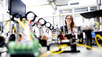
My focus was on the development of more sophisticated spectrometers – the “brains” of an MRI scanner, which carry out all the control, data acquisition and processing functions – and also more compact and effective MRI scanners for preclinical (that is, non-human) research. Our most significant innovation has been the elimination of the liquid helium cooling system for high-field superconducting MRI systems. In order to make a high-strength magnet, the magnet coils need to carry high current. In practice, this can be achieved only by using superconducting wire, which must be operated at or below 4 K.
Traditionally, this very low temperature was achieved by immersing the magnet coils in a bath of liquid helium, but this is undesirable for several reasons. The world’s supply of helium is limited, which makes it expensive, and “wet” magnets (those that use liquid helium) also require bulky and expensive safety features to guard against damage from a helium gas leak (for example, due to heating when the magnet undergoes a “quench” and ceases to be superconducting). So the challenge presented to our development team was to develop a cooling system that did not require helium, but could reliably cool the entire magnet uniformly.
The development of the helium-free technology was carried out in collaboration with our magnet partners, who have experience of building small magnets for other physics applications. The new design incorporates superconducting magnet coils that are cooled by direct conduction with an “off the shelf” cryocooler fridge unit. These systems have an up-front cost about half that of a traditional magnet, and they are compact and relatively lightweight (350 kg instead of more than two tonnes). This means they can be housed in ordinary laboratories, where space is at a premium, and they do not require an emergency exhaust system. In addition, regular top-ups of helium are no longer necessary.
Our scanners are used by university research departments and industrial firms all over the world
The other significant innovation was making it possible to perform other types of imaging (such as PET, SPECT and CT imaging) either in series or in parallel with the MRI imaging. This generates a fusion of complementary images and data and saves significant amounts of research time. Our scanners are used by university research departments and industrial firms (mainly pharmaceutical companies) all over the world, and their main application is imaging small animals. Being able to conduct many types of imaging in one unit is very helpful for these users, since it eliminates the need to move sedated animals around between imaging systems that, previously, may have been located in different laboratories.
Skills needed
We began shipping the compact, high-powered cryogen-free MRI scanner in 2012, and since then, our revenue has grown by more than 50%. In 2016 MR Solutions received the Queen’s Award for Enterprise (Innovation) at Buckingham Palace in recognition of our technical innovations.
At the moment, the biggest challenge we face is the availability of skilled staff here in the UK. The country’s manufacturing base has been eroded, and it’s hard to find experienced engineers, manufacturing managers and MRI physicists. Many overseas students do PhDs in MRI in the UK, but they don’t have the right to work here after they graduate and getting work permits for them is challenging. Also, the skills we need are often different from what the modern MRI research environment provides. A lot of students work on commercial clinical scanners for their PhDs, but we need people who can take a piece of equipment apart and rebuild it.
We do train people with an MRI physics background in these “hard skills”, though, and at the end of 2016, we were able to bring our magnet production closer to home, with a new magnet factory in Abingdon, Oxfordshire. This makes it possible to control quality and production, and to continue developing our technologies. So while it’s not as easy to make breakthroughs now as it was in the early days of MRI, I hope someday that we will see these compact magnets used in a clinical setting to treat patients.