Researchers in the US have improved the resolution for imaging the human eye by a third. This has enabled them to visualize the mosaic of rod and cone photoreceptors at the back of the eye in greater detail than ever before and allows the assessment of individual photoreceptors. The new imaging technique could facilitate earlier detection of diseases such as age-related macular degeneration and enhance the monitoring of treatments for eye diseases, the team claims.
Age-related macular degeneration is a progressive eye disease and a leading cause of sight loss. People with age-related macular degeneration lose the ability to see fine detail in their central vision, as the cells at the centre of their retina become damaged. Early diagnosis and treatment are crucial to preserve sight, in macular degeneration and other eye diseases.
“The goal of our research is to discern disease-related changes at the cellular level over time, possibly enabling much earlier detection of disease,” says Johnny Tam, at the National Eye Institute in Maryland. As well as improving the monitoring of degenerative changes in retinal tissue, enhanced image resolution could also help doctors see whether treatments for eye diseases are working and assist with the development and assessment of new therapies.
Imaging the eye is challenging. The light-distorting properties of parts of the eye like the lens and cornea reduce image resolution. Then there’s the diffraction limit of light to consider, with most conventional techniques for imaging beyond the diffraction limit using too much light to be safe for the eyes. Current retinal imaging techniques that use near-infrared light have a resolution of around 2−3 µm, while the smallest rod and cone photoreceptors range from 1–3 µm in diameter.
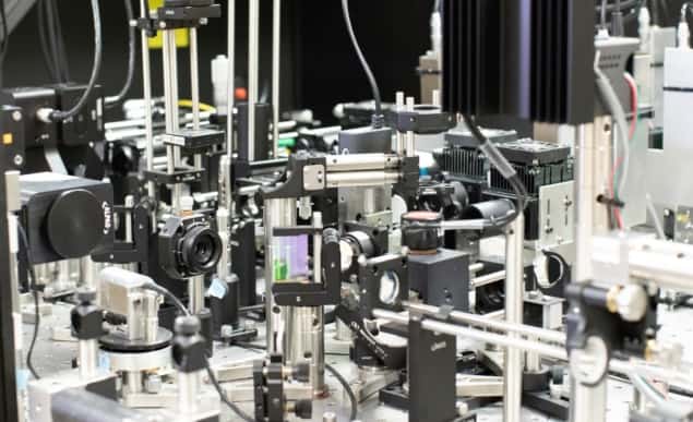
To improve resolution beyond the diffraction limit, and allow these individual cells to be distinguished, Tam and colleagues combined two imaging techniques: annular pupil illumination and sub-Airy disk confocal detection. By combining these two approaches, the researchers improved upon a conventional retinal imaging technique known as adaptive optics scanning light ophthalmoscopy, which uses deformable mirrors and computational methods to correct for optical imperfections of the eye in real time.
Annular pupil illumination generates a hollow beam of light. This improves the transverse resolution across the mosaic of photoreceptors, but reduces depth resolution. However, the researchers then used a very small pinhole – known as a sub-Airy disk – to block the light coming back from the eye, which allowed them to regain the depth resolution.
“One might think that more light is needed to get a better image, but we demonstrate that we can improve resolution by strategically blocking light in various locations within our instrument,” explains Tam. “This approach reduces the overall power of light delivered to the eye, making it ideal for live imaging applications.”
The researchers tested their technique on five adults with no sign of eye disease. Describing their research in Optica, they claim that this approach improved resolution across the photoreceptor mosaic by 33% and depth resolution by 13%, compared with conventional adaptive optics scanning light ophthalmoscopy. This allowed them to reveal subcellular features of photoreceptors that were not clearly visible with previous techniques.

“The ability to noninvasively image photoreceptors with subcellular resolution can be used to track how individual cells change over time,” says Tam. “For example, watching a cell begin to degenerate, and then possibly recover, will be an important advance for testing new treatments to prevent blindness.”
The researchers say that this clear improvement in the ability to obtain higher resolution images of the retina could help answer fundamental questions about photoreceptor health. They add that their technique provides a straightforward method of achieving sub-diffraction limited resolution in point-scanning-based microscopy and imaging approaches. This could enable routine sub-diffraction imaging of cells in the human body, they state, and be useful in other applications where it is important to image with low levels of light.


